Comparative Study
. 2011;13(5):R171.
doi: 10.1186/ar3494. Epub 2011 Oct 21.

An organ culture system to model early degenerative changes of the intervertebral disc

Affiliations
Comparative Study

An organ culture system to model early degenerative changes of the intervertebral disc

Ravi K Ponnappan et al. Arthritis Res Ther. 2011.

Abstract

Introduction: Back pain, a significant source of morbidity in our society, is related to the degenerative changes of the intervertebral disc. At present, the treatment of disc disease consists of therapies that are aimed at symptomatic relief. This shortcoming stems in large part from our lack of understanding of the biochemical and molecular events that drive the disease process. The goal of this study is to develop a model of early disc degeneration using an organ culture. This approach is based on our previous studies that indicate that organ culture closely models molecular events that occur in vivo in an ex vivo setting.

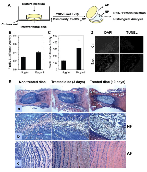
Methods: To mimic a degenerative insult, discs were cultured under low oxygen tension in the presence of TNF-α, IL-1β and serum limiting conditions.

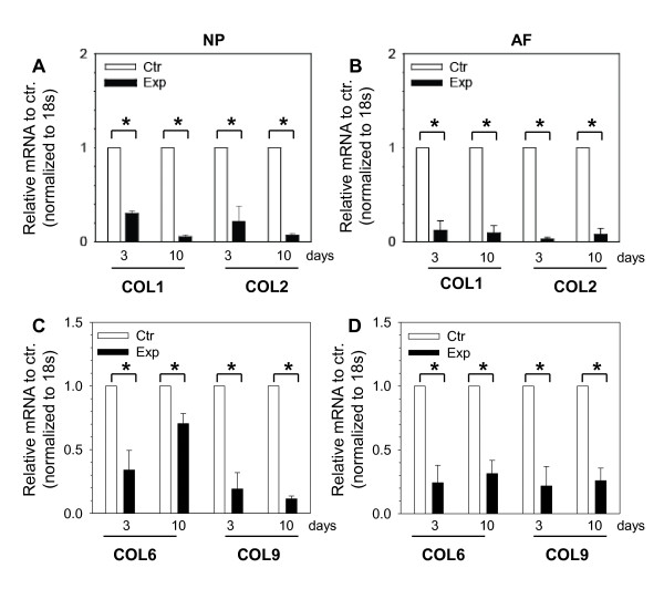
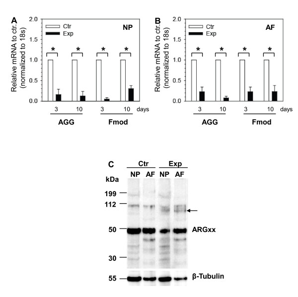
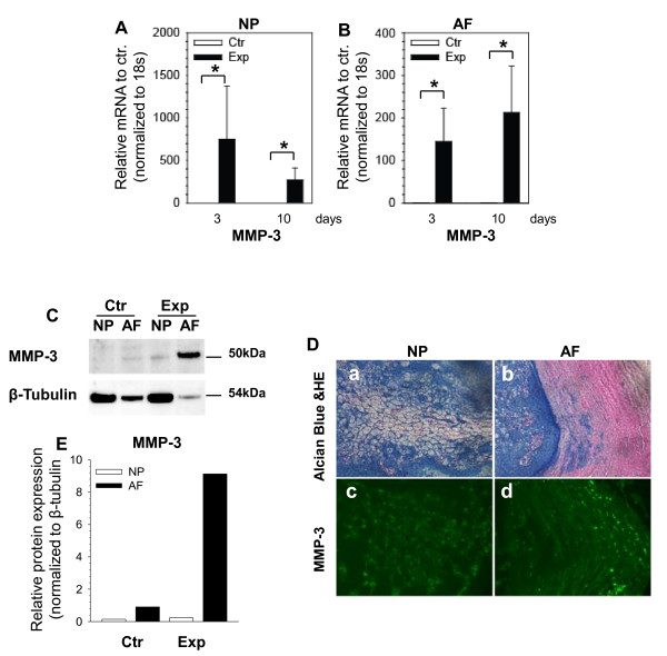
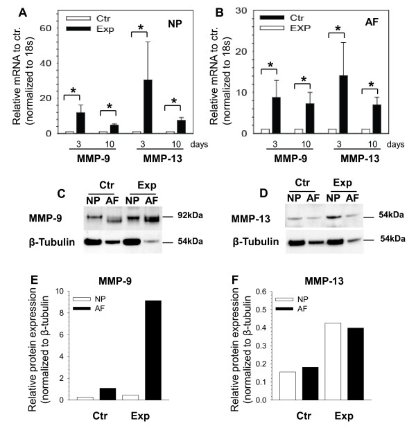
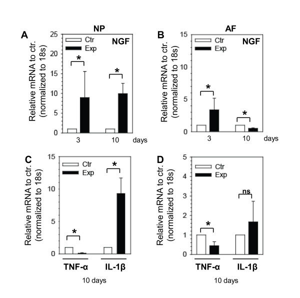
Results: Treatment resulted in compromised cell survival and changes in cellular morphology reminiscent of degeneration. There was strong suppression in the expression of matrix proteins including collagen types 1, 2, 6 and 9, proteoglycans, aggrecan and fibromodulin. Moreover, a strong induction in expression of catabolic matrix metalloproteinases (MMP) 3, 9 and 13 with a concomitant increase in aggrecan degradation was seen. An inductive effect on NGF expression was also noticed. Although similar, nucleus pulposus and annulus fibrosus tissues showed some differences in their response to the treatment.

Conclusions: Results of this study show that perturbations in microenvironmental factors result in anatomical and gene expression change within the intervertebral disc that may ultimately compromise cell function and induce pathological deficits. This system would be a valuable screening tool to investigate interventional strategies aimed at restoring disc cell function.


Figures

Figure 1
Figure 1
Establishment of organ culture model of disc degeneration. (A) Schematic showing disc organ culture setup. Culturing discs under low oxygen partial pressure (pO2) in hyperosmolar, nutritionally limiting media and exposure to proinflammatory cytokines mimic molecular changes characteristic of early disc degeneration. NP, nucleus pulposus; AF, annulus fibrosus; FBS, fetal bovine serum. (B,C) Measurement of luciferase diffusion in nucleus pulposus (NP) tissue after 24 hours. Both Firefly (61 kDa) and Renilla (38 KDa) luciferase diffuse in the tissue in a concentration-dependent fashion. Results are shown as the mean ± standard error for three independent experiments performed in triplicate. *P <0.05. (D) Histological sections were stained for apoptotic cells. Tumor necrosis factor-alpha (TNF-α)-treated and interleukin-1beta (IL-1β)-treated intervertebral discs demonstrated significantly higher numbers of apoptosis-positive cells (d) than the untreated control group (b) after 3 days in culture (DAPI: a and c). Ctr., control group; DAPI, 4'-6-diamidino-2-phenylindole; Exp., experimental group; TUNEL, terminal deoxynucleotidyl transferase-mediated dUTP-biotin nick end-labeling. (E) Histological analysis of disc after treatment. Sections are stained with Alcian blue and hematoxylin-and-eosin counter staining and photographed with a low-magnification (4×) lens (a). Panels (b) and (c) were examined under high magnification (20×). Aberrant cellular hypertrophy and matrix degradation of NP tissue in treated disc were observed after 3 and 10 days. Representative findings of three separate experiments (n = 3 discs/group per experiment) are shown.
Figure 2
Figure 2
Collagen gene expression is suppressed in organ culture model of disc degeneration. Expression of important disc matrix collagens decreases with tumor necrosis factor-alpha, interleukin-1beta, and limited nutrition. Expression of type 1 collagen (COL1) and type 2 collagen (COL 2) is significantly suppressed in (A) nucleus pulposus (NP) and (B) annulus fibrosus (AF) tissues of treated discs at 3 and 10 days. Note a significant decrease in type 6 collagen (COL 6) after the treatment in (C) NP and (D) AF. Type 9 collagen (COL 9) expression is decreased only in AF after 10 days of treatment (C,D). Results are shown as the mean ± standard error for three independent experiments (n = 6 discs/group per experiment). *P <0.05. Ctr., control group; Exp, experimental group.
Figure 3
Figure 3
Matrix gene expression is altered in organ culture model of disc degeneration. Real-time reverse transcription-polymerase chain reaction analysis of aggrecan (AGG) and fibromodulin after treatment. There is a significant downregulation in expression of AGG and fibromodulin (Fmod) mRNA in (A) nucleus pulposus (NP) and (B) annulus fibrosus (AF) tissues of treated discs. Results are shown as the mean ± standard error for three independent experiments. *P <0.05. (C) Presence of AGG fragments as analyzed by Western blotting in NP and AF tissue lysates after 3 days in culture. Arrows indicate the presence of additional AGG fragments recognized by anti-ARGxx antibody in the treated tissues. Representative findings of two independent experiments (n = 6 discs/group per experiment) are shown. Ctr., control group; Exp, experimental group.
Figure 4
Figure 4
Catabolic gene expression is upregulated by tumor necrosis factor-alpha, interleukin-1beta, and limited nutrition. Expression of matrix metalloproteinase 3 (MMP-3) mRNA in nucleus pulposus (NP) (A) and annulus fibrosus (AF) (B) tissues. Expression is significantly induced in both tissues after the treatment for 3 and 10 days. Results are shown as the mean ± standard error for three independent experiments (n = 6 discs/group per experiment). *P <0.05. (C) Western blot analysis of MMP-3 in NP and AF tissue lysates after 3 days in culture confirms the expression of MMP-3. (D) Representative photomicrographs of the immunohistochemical reactivity for MMP-3 in tissue sections from treated discs for 3 days (c,d). Sections were also stained with Alcian blue and hematoxylin and eosin (HE) for visualization of morphological details (a,b). Owing to extremely low signal intensity, data for untreated tissue sections are not shown. Original magnification: 20×. (E) The expression of MMP-3 tends to increase in the experimental group. Ctr., control group; Exp, experimental group.
Figure 5
Figure 5
Effect of tumor necrosis factor-alpha, interleukin-1beta, and limited nutrition on matrix metalloproteinase (MMP)-9 and MMP-13 expression. Expression analysis of MMP-9 and MMP-13 mRNA in nucleus pulposus (NP) (A) and annulus fibrosus (AF) (B) tissues after the treatment. Both MMP-9 and MMP-13 are significantly upregulated after the treatment. Results are shown as the mean ± standard error for three independent experiments (n = 6 discs/group per experiment). *P <0.05. Western blot analysis of MMP-9 (C) and MMP-13 (D) in NP and AF tissue lysates after 3 days in culture. The expression of MMP-9 (E) and MMP-13 (F) is increased in the treated samples. Ctr., control group; Exp, experimental group.
Figure 6
Figure 6
Tumor necrosis factor-alpha (TNF-α), interleukin-1beta (IL-1β), and limited nutrition causes changes in expression of genes associated with pain and inflammation. Nucleus pulposus (NP) (A) and annulus fibrosus (AF) (B) were analyzed at the indicated days for nerve growth factor (NGF) expression by using quantitative reverse transcription-polymerase chain reaction (RT-PCR). At 3 days of treatment, NGF is significantly increased in NP and AF. At 10 days after treatment, the expression of NGF in NP was significantly increased but in AF showed a lower expression compared with the control. Expression analysis of TNF-α and IL-1β genes showed decreased expression of TNF-α after treatment in NP (C) and AF (D) tissues. IL-1β showed an increase in expression compared with untreated control. Results are shown as the mean ± standard error for three independent experiments (n = 6 discs/group per experiment). *P <0.05. Ctr., control group; Exp, experimental group; ns, not significant.

Similar articles

Cited by

References

    1. Hoy D, March L, Brooks P, Woolf A, Blyth F, Vos T, Buchbinder R. Measuring the global burden of low back pain. Best Pract Res Clin Rheumatol. 2010;24:155–165. doi: 10.1016/j.berh.2009.11.002. - DOI - PubMed
    1. Dagenais S, Tricco AC, Haldeman S. Synthesis of recommendations for the assessment and management of low back pain from recent clinical practice guidelines. Spine J. 2010;10:514–529. doi: 10.1016/j.spinee.2010.03.032. - DOI - PubMed
    1. Boden SD, Davis DO, Dina TS, Patronas NJ, Wiesel SW. Abnormal magnetic-resonance scans of the lumbar spine in asymptomatic subjects. A prospective investigation. J Bone Joint Surg Am. 1990;72:403–408. - PubMed
    1. Pfirrmann CW, Metzdorf A, Zanetti M, Hodler J, Boos N. Magnetic resonance classification of lumbar intervertebral disc degeneration. Spine. 2001;26:1873–1878. doi: 10.1097/00007632-200109010-00011. - DOI - PubMed
    1. Thompson JP, Pearce RH, Schechter MT, Adams ME, Tsang IK, Bishop PB. Preliminary evaluation of a scheme for grading the gross morphology of the human intervertebral disc. Spine. 1990;15:411–415. doi: 10.1097/00007632-199005000-00012. - DOI - PubMed

Publication types

MeSH terms

LinkOut - more resources