. 2011 Oct 24:11:94.
doi: 10.1186/1471-2431-11-94.

Multi-species bacterial biofilm and intracellular infection in otitis media

Affiliations

Multi-species bacterial biofilm and intracellular infection in otitis media

Ruth B Thornton et al. BMC Pediatr. .

Abstract

Background: Bacteria which are metabolically active yet unable to be cultured and eradicated by antibiotic treatment are present in the middle ear effusion of children with chronic otitis media with effusion (COME) and recurrent acute otitis media (rAOM). These observations are suggestive of biofilm presence or intracellular sequestration of bacteria and may play a role in OM pathogenesis. The aim of this project is to provide evidence for the presence of otopathogenic bacteria intracellularly or within biofilm in the middle ear mucosa of children with COME or rAOM.

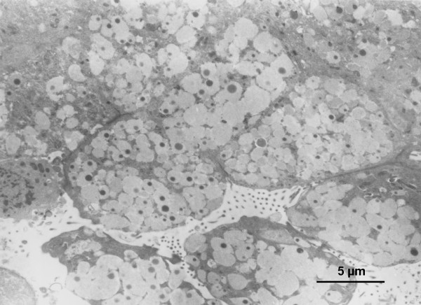
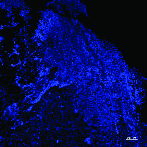
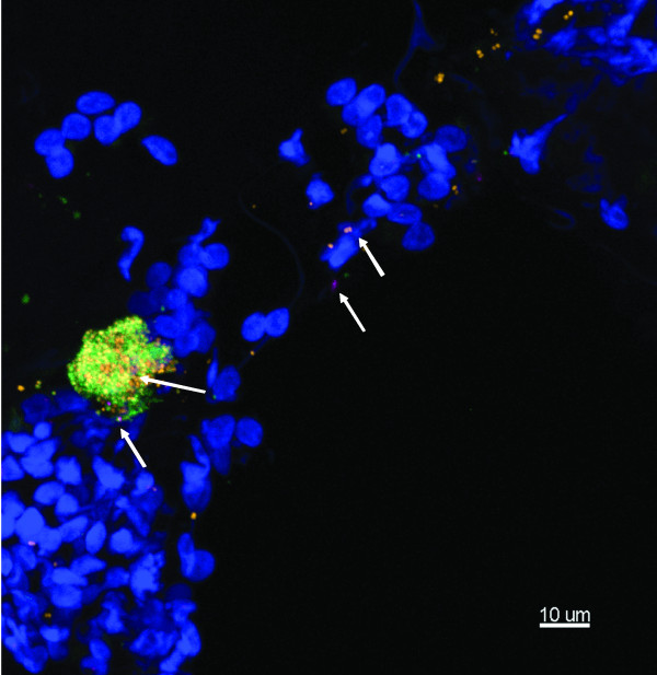
Methods: Middle ear mucosal biopsies from 20 children with COME or rAOM were examined for otopathogenic bacteria (either in biofilm or located intracellularly) using transmission electron microscopy (TEM) or species specific fluorescent in situ hybridisation (FISH) and confocal laser scanning microscopy (CLSM). One healthy control biopsy from a child undergoing cochlear implant surgery was also examined.

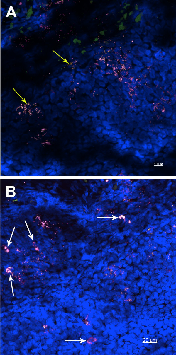
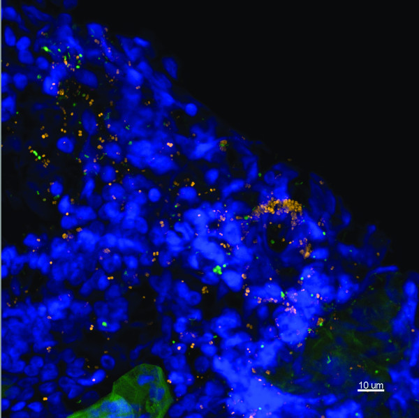
Results: No bacteria were observed in the healthy control sample. In 2 of the 3 biopsies imaged using TEM, bacteria were observed in mucus containing vacuoles within epithelial cells. Bacterial species within these could not be identified and biofilm was not observed. Using FISH with CLSM, bacteria were seen in 15 of the 17 otitis media mucosal specimens. In this group, 11 (65%) of the 17 middle ear mucosal biopsies showed evidence of bacterial biofilm and 12 demonstrated intracellular bacteria. 52% of biopsies were positive for both biofilm and intracellular bacteria. At least one otopathogen was identified in 13 of the 15 samples where bacteria were present. No differences were observed between biopsies from children with COME and those with rAOM.

Conclusion: Using FISH and CLSM, bacterial biofilm and intracellular infection with known otopathogens are demonstrated on/in the middle ear mucosa of children with COME and/or rAOM. While their role in disease pathogenesis remains to be determined, this previously undescribed infection pattern may help explain the ineffectiveness of current treatment strategies at preventing or resolving COME or rAOM.


Figures

Figure 1
Figure 1
Representative TEM of the middle ear mucosa from a child with a history of COME and recurrent AOM. This biopsy was from a 25 month old male, MEE was not collected. Image shows mucus-secreting epithelial cells (12-15 μm) overwhelmed with coccal bacteria in mucus containing vacuoles.
Figure 2
Figure 2
Representative image from the middle ear mucosal biopsy of a child having a cochlear implant who has no history of middle ear disease. Child 21. FISH -EUB338 (Yellow), S. pneumoniae (green), H. influenzae (pink) and Hoechst 33342 (nuclei stain - blue). This maximum intensity projection (Z = 20.5 μm) demonstrates the normal mucosal tissue with no evidence of bacteria. Scale bar = 50 μm.
Figure 3
Figure 3
Representative images of a mucosal biopsy from a child suffering with rAOM. Middle ear fluid culture and Ply PCR negative, showing biofilm and intracellular infection. Child 4. FISH: EUB338 (yellow), M. catarrhalis (green), H. influenzae (pink) and Hoechst 33342 (nuclei stain - Blue). A) H. influenzae is the predominant pathogen, with other non-identified bacteria also evident (yellow arrows). Maximum projection image (Z = 29 μm), scale bar = 10 μm. B) Intracellular H. influenzae are apparent throughout the tissue as nuclei associated bacterial clusters (white arrows), as well as in biofilm. Maximum projection image (Z = 40 μm), scale bar = 20 μm.
Figure 4
Figure 4
Representative image of a mucosal biopsy from a child suffering from rAOM and COME. MEE was not present. Child 14. FISH probes included EUB338 (yellow), S. pneumoniae (green), negative for H. influenzae (pink), Hoechst 33342 (nuclei stain - blue). This is a maximum intensity projection (Z = 39 μm) showing multispecies biofilm covering the mucosa. The biofilm is seen to consist of S. pneumoniae and other unidentified bacteria. These bacteria are also interspersed within the tissue. Scale bar = 10 μm.
Figure 5
Figure 5
Representative image of a mucosal biopsy from a child with a history of COME. No middle ear fluid was taken. Child 20. FISH - EUB338 (Yellow), M. catarrhalis (green), S. pneumoniae (pink) and Hoechst 33342 (nuclei stain - blue). Maximum intensity projection (Z = 19.5 μm) showing microcolony formation, mainly M. catarrhalis and S. pneumoniae (arrows) scattered throughout the mucosa. Scale bar = 10 μm.

References

    1. Park CW, Han JH, Jeong JH, Cho SH, Kang MJ, Tae K, Lee SH. Detection rates of bacteria in chronic otitis media with effusion in children. J Korean Med Sci. 2004;19(5):735–738. doi: 10.3346/jkms.2004.19.5.735. - DOI - PMC - PubMed
    1. Rayner MG, Zhang Y, Gorry MC, Chen Y, Post JC, Ehrlich GD. Evidence of bacterial metabolic activity in culture-negative otitis media with effusion. Jama. 1998;279(4):296–299. doi: 10.1001/jama.279.4.296. - DOI - PubMed
    1. Parsek MR, Singh PK. Bacterial biofilms: an emerging link to disease pathogenesis. Annu Rev Microbiol. 2003;57:677–701. doi: 10.1146/annurev.micro.57.030502.090720. - DOI - PubMed
    1. Burmolle M, Thomsen TR, Fazli M, Dige I, Christensen L, Homoe P, Tvede M, Nyvad B, Tolker-Nielsen T, Givskov M. et al.Biofilms in chronic infections - a matter of opportunity - monospecies biofilms in multispecies infections. FEMS Immunol Med Microbiol. 2010;59(3):324–336. - PubMed
    1. Ehrlich GD, Veeh R, Wang X, Costerton JW, Hayes JD, Hu FZ, Daigle BJ, Ehrlich MD, Post JC. Mucosal biofilm formation on middle-ear mucosa in the chinchilla model of otitis media. Jama. 2002;287(13):1710–1715. doi: 10.1001/jama.287.13.1710. - DOI - PubMed

Publication types