

doi: 10.1053/j.ajkd.2011.08.009.
Epub 2011 Oct 21.
CKD-mineral and bone disorder: core curriculum 2011
Affiliations
- PMID: 22018457
- PMCID: PMC3983665
- DOI: 10.1053/j.ajkd.2011.08.009
Item in Clipboard
CKD-mineral and bone disorder: core curriculum 2011
Am J Kidney Dis.
2011 Dec.
No abstract available
Figures
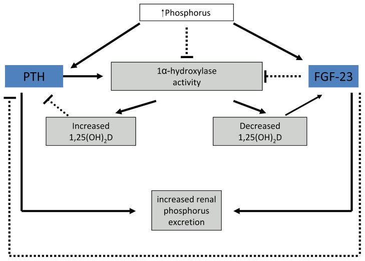
A solid line indicates stimulation; a dashed line indicates inhibition. Adapted with permission of Elsevier from Moe SM, Sprague SM. Mineral bone disorders in chronic kidney disease. In: Brenner and Rector’s The Kidney, 8th ed. Philadelphia, PA: WB Saunders Company; 2007:1784–1807.
Adapted with permission of Elsevier from Moe SM, Sprague SM. Mineral bone disorders in chronic kidney disease. In: Brenner and Rector’s The Kidney, 8th ed. Philadelphia, PA: WB Saunders Company; 2007:1784–1807.
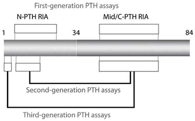
The intact PTH molecule is composed of 84 amino acids; different regions of the protein are targeted by first through third generation assays. Mid/C-PTH, mid/carboxyl terminus of PTH; N-PTH, amino terminus of PTH; RIA, radioimmunoassay. Reproduced with permission of Elsevier from Moe SM, Sprague SM. Mineral bone disorders in chronic kidney disease. In: Brenner and Rector’s The Kidney, 8th ed. Philadelphia, PA: WB Saunders Company; 2007:1784–1807.
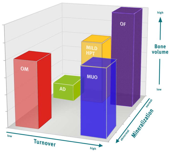
The TMV system provides more information than the previously used classification scheme. Each axis represents one of the descriptors in the TMV classification: turnover (from low to high), mineralization (from normal to abnormal), and bone volume (from low to high). Individual patient parameters can be plotted on the graph, or means and ranges of grouped data can be shown. For example, many patients with renal osteodystrophy cluster in areas shown by the bars. The red bar (OM, osteomalacia) was previously described as low-turnover bone with abnormal mineralization. The bone volume may be low to medium, depending on the severity and duration of the process and other factors that affect bone. The green bar (AD, adynamic bone disease) was previously described as low-turnover bone with normal mineralization, and the bone volume in this example is at the lower end of the spectrum, but other patients with normal mineralization and low turnover will have normal bone volume. The yellow bar (mild HPT, mild hyperparathyroid-related bone disease) and purple bar (OF, osteitis fibrosa or advanced hyperparathyroid-related bone disease) were previously considered distinct categories, but in actuality represent a range of abnormalities along a continuum of medium to high turnover, and any bone volume depending on the duration of the disease process. Finally, the blue bar (MUO, mixed uremic osteodystrophy) is variably defined internationally. In the present graph, it is depicted as high-turnover, normal bone volume, with abnormal mineralization. In summary, the TMV classification system more precisely describes the range of pathologic abnormalities that can occur in patients with CKD. Reproduced with permission of Nature Publishing Group from Figure 1 in Moe et al. Kidney Int. 2006;69(11):1945–53.
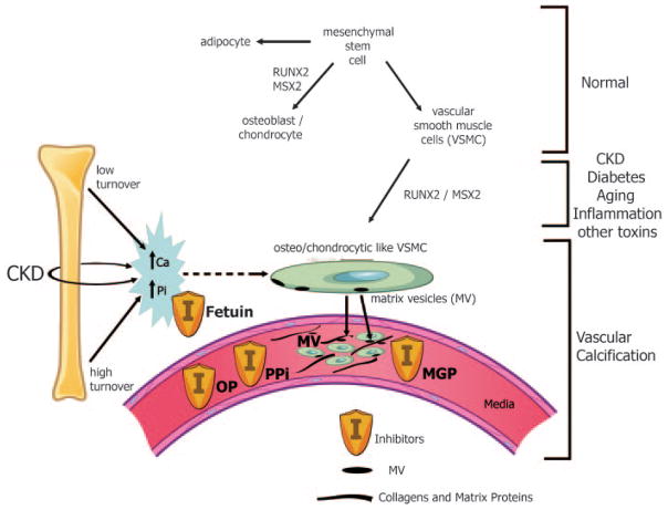
Normally, mesenchymal stem cells differentiate to adipocytes, osteoblasts, chondrocytes, and vascular smooth muscle cells (VSMC). In the setting of CKD, diabetes, aging, inflammation, and multiple other toxins, these VSMC can dedifferentiate or transform into osteo/chondrocytic-like cells by upregulation of transcription factors such as RUNX-2 and MSX2. These transcription factors are critical for normal bone development and thus their upregulation in VSMC is indicative of a phenotypic switch. These osteo/chondrocytic-like VSMC then become calcified in a process similar to bone formation. These cells lay down collagen and noncollagenous proteins in the intima or media and incorporate calcium and phosphorus into matrix vesicles to initiate mineralization and further mineralize into hydroxyapatite. The overall positive calcium and phosphorus balance of most dialysis patients feeds both the cellular transformation and the generation of matrix vesicles. In addition, the extremes of bone turnover in chronic kidney disease (low and high or adynamic and hyperparathyroid bone, respectively) will increase the available calcium and phosphorus by altering the bone content of these minerals. Ultimately, whether an artery calcifies or not depends on the strength of the army of inhibitors (I) standing by in the circulation (fetuin A) and in the arteries (PPI = pyrophosphate, MGP = matrix Gla protein, and OP = osteopontin as examples). Reproduced with permission of the American Society of Nephrology from Figure 1 in Moe et al. J Am Soc Nephrol. 2008;19:213–216.
Similar articles
-
Effectiveness and cost-efficacy of phosphate binders in hemodialysis.Ann Nutr Metab. 2011 Oct;58(4):315-9. doi: 10.1159/000331988. Epub 2011 Oct 7. Ann Nutr Metab. 2011. PMID: 21986491
-
Parathyroid function in chronic kidney disease: role of FGF23-Klotho axis.Contrib Nephrol. 2013;180:110-23. doi: 10.1159/000346791. Epub 2013 May 3. Contrib Nephrol. 2013. PMID: 23652554 Review.
-
[Control of phosphorus and treatment with vitamin D in chronic kidney disease prior to the start of dialysis].Nefrologia. 2008;28 Suppl 5:39-45. Nefrologia. 2008. PMID: 18847419 Review. Spanish.
-
Chronic kidney disease: Phosphate binder therapy--cracks in the tower of strength?Nat Rev Nephrol. 2012 Nov;8(11):615-6. doi: 10.1038/nrneph.2012.219. Epub 2012 Oct 9. Nat Rev Nephrol. 2012. PMID: 23045230 No abstract available.
-
Effects of phosphate binders in moderate CKD.J Am Soc Nephrol. 2012 Aug;23(8):1407-15. doi: 10.1681/ASN.2012030223. Epub 2012 Jul 19. J Am Soc Nephrol. 2012. PMID: 22822075 Free PMC article. Clinical Trial.
Cited by
-
Intestinal Phosphorus Absorption in Chronic Kidney Disease.Nutrients. 2018 Sep 23;10(10):1364. doi: 10.3390/nu10101364. Nutrients. 2018. PMID: 30249044 Free PMC article. Review.
-
Bone-eating kidney disease.SAGE Open Med Case Rep. 2017 Dec 5;5:2050313X17744983. doi: 10.1177/2050313X17744983. eCollection 2017. SAGE Open Med Case Rep. 2017. PMID: 29238578 Free PMC article.
-
Fibroblast growth factor 23 and Klotho: physiology and pathophysiology of an endocrine network of mineral metabolism.Annu Rev Physiol. 2013;75:503-33. doi: 10.1146/annurev-physiol-030212-183727. Annu Rev Physiol. 2013. PMID: 23398153 Free PMC article. Review.
-
Coronary artery calcification in patients with advanced chronic kidney disease.BMC Cardiovasc Disord. 2022 Oct 29;22(1):453. doi: 10.1186/s12872-022-02879-0. BMC Cardiovasc Disord. 2022. PMID: 36309659 Free PMC article.
-
First-in-Human Phase I Study of the Novel Injectable Calcimimetic Agent Upacicalcet in Healthy Adult Japanese Participants.Drugs R D. 2022 Jun;22(2):131-140. doi: 10.1007/s40268-022-00385-4. Epub 2022 Mar 25. Drugs R D. 2022. PMID: 35338469 Free PMC article. Clinical Trial.
References
-
- Moe S, Drueke T, Cunningham J, et al. Definition, evaluation, and classification of renal osteodystrophy: a position statement from Kidney Disease: Improving Global Outcomes (KDIGO) Kidney Int. 2006 Jun;69(11):1945–1953. - PubMed
-
- KDIGO clinical practice guideline for the diagnosis, evaluation, prevention, and treatment of Chronic Kidney Disease-Mineral and Bone Disorder (CKD-MBD) Kidney Int Suppl. 2009 Aug;113:S1–S130. - PubMed
-
- Moe SM, Drueke T, Lameire N, Eknoyan G. Chronic kidney disease-mineral-bone disorder: a new paradigm. Adv Chronic Kidney Dis. 2007 Jan;14(1):3–12. - PubMed
-
- Moe SM, Sprague SM. Brenner and Rector’s The Kidney. 8. WB Saunders Company; Philadelphia, PA: 2007. Chapter 52: Mineral bone disorders in chronic kidney disease; pp. 1784–1807.
-
- Moe SM. In: Chapter 8: Chronic Kidney Disease, Dialysis, and Transplantation. 3. Himmelfarb J, Sayegh M, editors. Elsevier Saunders; Philadelphia: 2010. pp. 98–114.
MeSH terms
Substances
Grants and funding
LinkOut - more resources
Full Text Sources
Medical
