. 2011 Oct 28;35(4):633-46.
doi: 10.1016/j.immuni.2011.08.016. Epub 2011 Oct 20.

Differential expression of Ly6C and T-bet distinguish effector and memory Th1 CD4(+) cell properties during viral infection

Affiliations

Differential expression of Ly6C and T-bet distinguish effector and memory Th1 CD4(+) cell properties during viral infection

Heather D Marshall et al. Immunity. .

Abstract

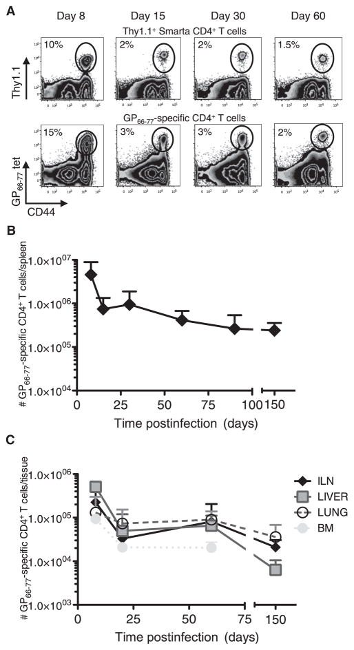
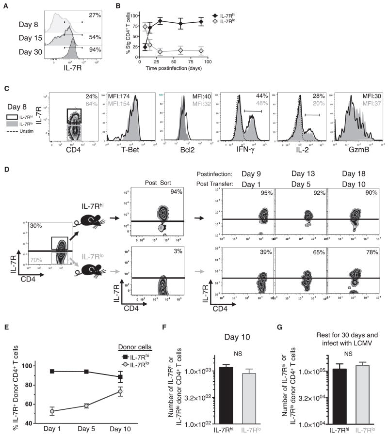
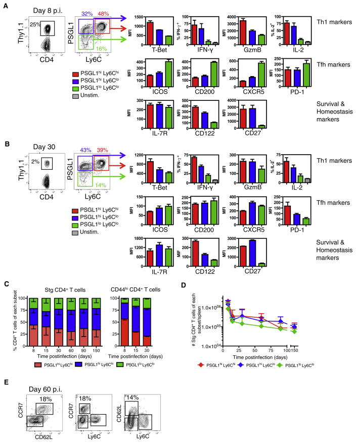
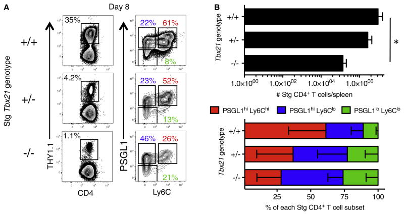
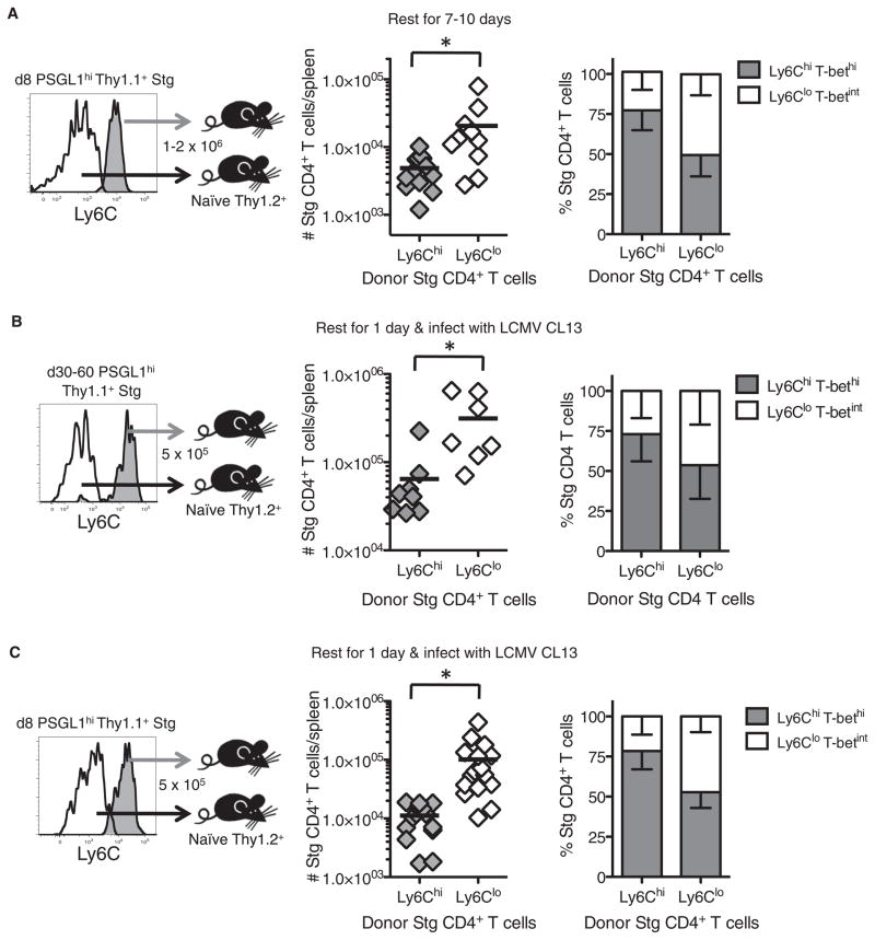
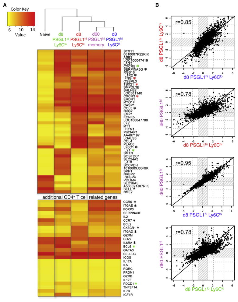
CD4(+) T cells differentiate into multiple effector types, but it is unclear how they form memory T cells during infection in vivo. Profiling virus-specific CD4(+) T cells revealed that effector cells with T helper 1 (Th1) or T follicular helper (Tfh) cell characteristics differentiated into memory cells, although expression of Tfh cell markers declined over time. In contrast to virus-specific effector CD8(+) T cells, increased IL-7R expression was not a reliable marker of CD4(+) memory precursor cells. However, decreased Ly6C and T-bet (Tbx21) expression distinguished a subset of Th1 cells that displayed greater longevity and proliferative responses to secondary infection. Moreover, the gene expression profile of Ly6C(lo)T-bet(int) Th1 effector cells was virtually identical to mature memory CD4(+) T cells, indicating early maturation of memory CD4(+) T cell features in this subset during acute viral infection. This study provides a framework for memory CD4(+) T cell development after acute viral infection.


Figures

Figure 1
Figure 1. Kinetics of the LCMV-Specific GP66–77 Stg and Polyclonal CD4+ T Cell Responses
(A) Stg chimeric mice (top) or B6 mice (bottom) were infected with LCMV Armstrong, and 8, 15, 30, and 60 days p.i., splenocytes were analyzed for Thy1.1+ or GP66–77 tetramer+ CD4+ T cells. (B and C) The number of GP66–77-specific CD4+ T cells in the spleen (B), inguinal lymph nodes, liver, lung, and bone marrow (C) was determined at various time points p.i. Cumulative numbers of tetramer and Stg TCR transgenic cells from 9–7 mice/time point from more than 20 independent experiments are graphed + SD. See also Figure S1.
Figure 2
Figure 2. IL-7R Expression Does Not Mark Memory Precursor Effector CD4+ T Cells
(A and B) Frequency of IL-7R on virus-specific CD4+ T cells was determined at various time points p.i. and shown in histogram plots (A) and in line graph ± SD (B). (C) Expression of the indicated proteins was compared between IL-7Rhi (black line) and IL-7Rlo (gray shaded) Stg CD4+ T cells at day 8 p.i. and shown in overlapping histograms. (D–F) IL-7Rhi (black) and IL-7Rlo (gray) Stg CD4+ T cells were purified by FACS 8 days p.i. and equal numbers were transferred into infection-matched congenic recipients. Donor cells in the blood were analyzed for IL-7R expression over 10 days posttransfer and the frequency of IL-7Rhi donor T cells are shown in representative FACS plots (D) or cumulative line graph ± SD (E). (F) Bar graph shows the absolute number of donor cells + SD recovered 10 days posttransfer. (G) As in (D) above, except the donor IL-7Rhi and IL-7Rlo effector T cells were parked for 30 days in naive congenic recipients that were subsequently infected with LCMV. Seven days after secondary infection, the number of donor Stg CD4+ T cells in the spleen + SD was measured. Data are cumulative of three independent experiments. NS, not statistically significant.
Figure 3
Figure 3. Formation of Distinct LCMV-Specific Effector CD4+ T Cells Subsets
(A and B) Stg chimeric mice were infected with LCMV and 8 (A) or ~30 (B) days p.i. splenocytes were analyzed by flow cytometry for expression of the indicated proteins. PSGL1hiLy6Chi (red), PSGL1hiLy6Clo (blue), and PSGL1loLy6Clo (green) Stg CD4+ T cells were gated and the average MFI + SD for the various proteins are shown in bar graphs. CD4+ T cells were also stimulated with GP61–80 peptide in vitro and the average frequency of IFN-γ+ and IL-2+ Stg T cells + SD are shown. Data shown are representative of three independent experiments (n = 3 per group). (C) Stacked bar graph of Stg CD4 T cells (left) or CD44hi polyclonal CD4+ T cells (right) – SD in each subset after LCMV infection. (D) Total number of Stg CD4+ T cells + SD of each subset during LCMV infection. Cumulative data from 9–37 mice/time point from more than 20 independent experiments are graphed. (E) The expression of CD62L and CCR7 on Ly6Chi and Ly6Clo Stg CD4+ T cells at day 60 p.i. is shown in FACS plots. See also Figure S2.
Figure 4
Figure 4. T-bet-Dependent Formation of Antiviral CD4+ T Cell Subsets
1 × 104 Tbx21+/+, Tbx21+/−, or Tbx21−/− Stg CD4+ T cells were adoptively transferred into three groups of mice and then infected with LCMV Armstrong. Eight days p.i. the frequency of Stg cells and expression of PSGL1 and Ly6C was analyzed. Representative examples in FACS plots (A) and cumulative numbers of donor Stg CD4+ T cells + SD (B) and frequencies – SD of the different effector CD4+ T cell subsets from three independent experiments. *p < 0.05.
Figure 5
Figure 5. Enhanced Memory Cell Properties in PSGL1hiLy6Clo Effector CD4+ T Cells
(A) PSGL1hiLy6Chi or PSGL1hiLy6Clo Stg CD4+ T cells were purified 8 days p.i. and adoptively transferred at equal numbers (~1.5 × 10⁶) into naive congenic recipients. 7–10 days later, the number of donor Stg CD4+ T cells remaining in the spleen and their phenotypes – SD were determined. Cumulative data from six independent experiments is shown; black line represents the mean. (B and C) 5 × 10⁵ PSGL1hiLy6Chi or PSGL1hiLy6Clo Stg CD4+ T cells were purified 30–60 (B) or 8 (C) days p.i. and transferred into mice that were subsequently infected with LCMV clone 13. Six days later, the numbers of splenic donor Stg CD4+ T cells and their phenotypes were determined – SD. Cumulative data from three (B) or five (C) independent experiments are shown. *p < 0.05.
Figure 6
Figure 6. Gene Expression Profiles of PSGL1hiLy6Clo Effector CD4+ T Cells and Memory CD4+ T Cells Are Highly Similar
Three independent Stg CD4+ T cell samples containing naive, day 8 PSGL1loLy6Clo, day 8 PSGL1hiLy6Chi, day 8 PSGL1hiLy6Clo, or day 60 PSGL1hi memory CD4+ T cells were purified by FACS, and whole-genome mRNA expression profiles were characterized with Illumina microarrays. See also Table S1. (A) Clustering analysis and heatmap of expression values showing the log2 transformed expression intensity of any gene with a 32-fold change or higher over naive (q < 0.0001). Genes marked by an asterisk are mentioned in the Results or Discussion. (B) The overall similarity in gene expression between the different effector and memory CD4+ T cell samples was examined, in a pairwise manner, by identifying all the genes that were differentially expressed in any sample relative to naive CD4+ T cells (greater than 2-fold change and q < 0.05) and then plotting their relative intensities in each respective sample compared to the naive population.

Comment in

References

    1. Bamezai A. Mouse Ly-6 proteins and their extended family: markers of cell differentiation and regulators of cell signaling. Arch Immunol Ther Exp (Warsz) 2004;52:255–266. - PubMed
    1. Blander JM, Sant’Angelo DB, Bottomly K, Janeway CA., Jr Alteration at a single amino acid residue in the T cell receptor alpha chain complementarity determining region 2 changes the differentiation of naive CD4 T cells in response to antigen from T helper cell type 1 (Th1) to Th2. J Exp Med. 2000;191:2065–2074. - PMC - PubMed
    1. Chandele A, Joshi NS, Zhu J, Paul WE, Leonard WJ, Kaech SM. Formation of IL-7Ralphahigh and IL-7Ralphalow CD8 T cells during infection is regulated by the opposing functions of GABPalpha and Gfi-1. J Immunol. 2008;180:5309–5319. - PMC - PubMed
    1. De Riva A, Bourgeois C, Kassiotis G, Stockinger B. Noncognate interaction with MHC class II molecules is essential for maintenance of T cell metabolism to establish optimal memory CD4 T cell function. J Immunol. 2007;178:5488–5495. - PubMed
    1. Fazilleau N, Eisenbraun MD, Malherbe L, Ebright JN, Pogue-Caley RR, McHeyzer-Williams LJ, McHeyzer-Williams MG. Lymphoid reservoirs of antigen-specific memory T helper cells. Nat Immunol. 2007;8:753–761. - PubMed

Publication types

Associated data