Comparative Study
. 2011 Nov 24:1425:1-12.
doi: 10.1016/j.brainres.2011.09.045. Epub 2011 Sep 29.

Effects of inhibitory amino acids on expression of GABAA Rα and glycine Rα1 in hypoxic rat cortical neurons during development

Affiliations
Comparative Study

Effects of inhibitory amino acids on expression of GABAA Rα and glycine Rα1 in hypoxic rat cortical neurons during development

Hong Qian et al. Brain Res. .

Abstract

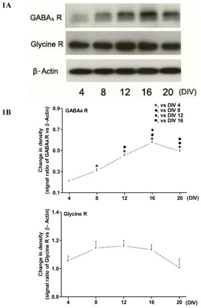
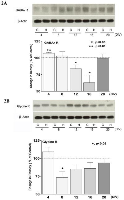
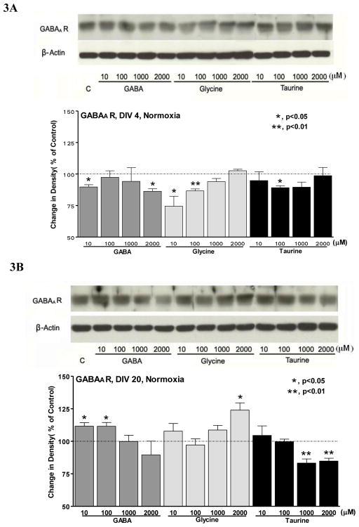
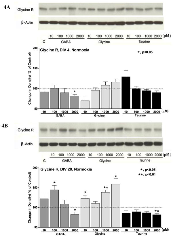
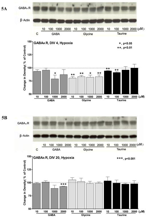
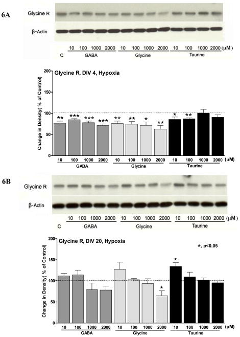
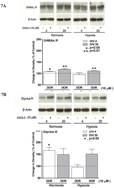
Recent studies suggest that GABA and glycine are protective to mature but toxic to immature cortical neurons during prolonged hypoxia. Since the action of these inhibitory amino acids is mediated by GABA and glycine receptors, the expression of these receptors is a critical factor in determining neuronal response to GABA(A) and glycine in hypoxia. Therefore, we asked whether in rat cortical neurons, 1) hypoxia alters the expression of the GABA and glycine receptors; 2) inhibitory amino acids change the course of GABA and glycine receptor expression; and 3) there are any differences between the immature and mature neurons. In cultured rat cortical neurons from day 4 (four days in vitro or DIV 4) to day 20 (DIV 20), we observed that 1) GABA(A)Rα and GlyRα1 underwent differential changes in expression during the development in vitro; 2) hypoxia for 3 days decreased GABA(A)Rα and GlyRα1 density in the neurons in-between DIV 4 and DIV 20, but did not induce a major change in immature (DIV 4) and mature (DIV 20) neurons; 3) during normoxia GABA, glycine and taurine decreased GABA(A)Rα and GlyRα1 density in the immature neurons, but had a tendency to increase the density in the mature neurons, except for taurine; 4) under hypoxia, all these amino acids decreased GABA(A)Rα and GlyRα1 density in most groups of the immature neurons with a slight effect on the mature neurons; and 5) δ-opioid receptor activation with DADLE increased GABA(A)Rα and GlyRα1 density in both the immature and mature neurons under normoxia and in the mature neurons under hypoxic condition. These data suggest that inhibitory amino acids differentially regulate the expression of GABA(A) and glycine receptors in rat cortical neurons under normoxic and hypoxic conditions with major differences between the immature and mature neurons.


Figures

Fig. 1
Fig. 1. Major differences in GABAA and glycine receptor expression during neuronal development
1A, Representative images of Western blot analysis. GABAA and glycine receptors were detected at different culture days from DIV 4 to 20 (n=4). 1B, Summary of changes in GABAA and glycine receptor levels with increase in days in culture (n=4). The asterisks (*), dagger (◆), ring (●) and square (■) indicate significant differences as compared to DIV 4, DIV 8, DIV 12 and DIV 16, respectively (P<0.05). Note that GABAA receptor density in immature (e.g., DIV 4) neurons was less than one-third of the mature level. The density increased with culture days, reaching the peak level at DIV 16 and then slightly decreasing to the level at DIV 20. In sharp contrast, glycine receptor had a high density in DIV-4 neurons. There was no significant change in the receptor density from DIV 4 to 20.
Fig. 2
Fig. 2. Hypoxic regulation of GABAA and glycine receptor expression during neuronal development
The control level in each group was expressed as 100% (dashed line). 2A, Effect of hypoxia on GABAA receptor expression. Cultured cortical neurons were treated or left untreated (normoxic control) with hypoxia for 72 hours. Top, representative blots at different culture ages. Bottom, statistical summary (n=4) with asterisks indicate a significant difference from the control. Note that hypoxia slightly increased GABAA receptor density in DIV-4 neurons, while it had no appreciable effect on DIV-8 neurons. The same hypoxic stress, however, significantly decreased GABAA receptor density in later ages, i.e., DIV 12 and 16 though it did not affect DIV-20 neurons. 2B, Effect of hypoxia on glycine receptor expression. Top, representative blots at different culture ages. Bottom, statistical data (n=4) with asterisks indicating a significant difference from the control. Note that hypoxia tended to increase glycine receptor expression in DIV-4 neurons, but significantly reduced the expression in DIV-8 neurons.
Fig. 3
Fig. 3. Effects of inhibitory neurotransmitters on GABAA receptor expression of immature and mature cortical neurons in normoxia, c, control
The control level in each group was expressed as 100% (dashed line). 3A, Effect of inhibitory neurotransmitters on GABAA receptor expression in immature (DIV-4) neurons. Top, representative blots. Bottom, statistical analysis of the signal density with the asterisks representing significant differences as compared with the control (n=3). Note that in DIV-4 neurons, GABA, glycine and taurine tended to attenuate GABAA receptor expression. 3B, Effects of inhibitory neurotransmitters on GABAA receptor expression in mature (DIV-20) neurons. Top, representative blots. Bottom, statistical analysis of the band density. The asterisks represent significant differences as compared with the control (n=3). Note that GABAA receptor density was significantly decreased by highest concentration of GABA (2000 μM), and highest concentration of glycine, which was different from changes in DIV-4 neurons. In contrast, taurine showed a tendency similar to that seen in DIV-4 neurons with higher concentrations (1000 μM and 2000 μM) significantly decreased GABAA receptor density.
Fig. 4
Fig. 4. Effects of inhibitory neurotransmitters on glycine receptor expression of immature and mature cortical neurons in normoxia. c, control
The control level in each group was expressed as 100% (dashed line). 4A, Effect of inhibitory neurotransmitters on glycine receptor expression in immature (DIV-4) neurons. Top, representative blots. Bottom, statistical analysis of the band density with the asterisks representing significant differences as compared with the control (n=3). Note that a high concentration (2000 μM) of GABA and a low concentration (10 μM) of glycine tended to decrease glycine receptor density, while taurine did not cause significant change in glycine receptor density though a low concentration (10 μM) tended to increase it. 4B, Effect of inhibitory neurotransmitters on glycine receptor expression in mature (DIV-20) neurons. Top, representative blots. Bottom, statistical analysis of the band density. The asterisks represent significant differences as compared with the control (n=3). Note that both GABA and glycine tended to increase glycine receptor density except the highest concentration (2000 μM) GABA, while taurine markedly decreased the glycine receptor density.
Fig. 5
Fig. 5. Effects of inhibitory neurotransmitters on GABAA receptor expression of immature and mature cortical neurons in hypoxia
c, control (hypoxia without inhibitory neurotransmitters). The control level in each group was expressed as 100% (dashed line). 5A, Effects of inhibitory neurotransmitters on GABAA receptor expression in DIV-4 neurons. Top, representative blots. Bottom, statistical analysis of the band density. The asterisks represent significant differences as compared with the control (n=3). Note that GABAA receptor density decreased in response to most concentrations of GABA, glycine and taurine in hypoxic condition. 5B, Effects of inhibitory neurotransmitters on GABAA receptor expression in DIV-20 neurons. Top, representative blots. Bottom, statistical analysis of the band signals with the asterisks representing significant differences as compared with the control (n=3). Note that except the high concentration GABA (2000 μM) that reduced GABAA receptor density, all other concentrations of inhibitory neurotransmitters had no significant effect on the receptor density.
Fig. 6
Fig. 6. Effects of inhibitory neurotransmitters on glycine receptor expression of immature and mature cortical neurons in hypoxia
c, hypoxic control (hypoxia without inhibitory neurotransmitters). The control level in each group was expressed as 100% (dashed line). 6A, Effects of inhibitory neurotransmitters on glycine receptor expression in DIV-4 neurons. Top, representative blots. Bottom, statistical analysis of the band density. The asterisks represent significant differences as compared with the control (n=4). Note that almost all concentrations of inhibitory neurotransmitters induced a decrease in glycine receptor expression. 6B, Effects of inhibitory neurotransmitters on glycine receptor expression in DIV-20 neurons. Top, representative blots. Bottom, statistical analysis of the band density. The asterisks represent significant differences as compared with the control (n=3). Note that most concentrations of inhibitory neurotransmitters did not cause significant change except the highest concentration (2000 μM) of glycine that significantly decreased the density and the lowest concentration (10 μM) of taurine that increased the density.
Fig. 7
Fig. 7. Effect of DOR activation (DOR) on GABAA and glycine receptor expression in normoxia and during hypoxia
7A, Effect of DOR activation on GABAA receptor expression. Top, representative Western blots. Bottom, statistical data (n=3) with asterisk indicating a significant difference from the control (no activation of DOR). The control level in each group was expressed as 100% (dashed line). Note that DOR activation with DADLE slightly increased GABAA receptor density in normoxia and hypoxia except DIV-4 neurons during hypoxia. 7B, Effect of DOR activation on glycine receptor expression. Top, representative blots. Bottom, statistical data (n=3) with the asterisk indicating significant difference from the control (no activation of DOR). Note that DOR-induced change in glycine receptor density was similar to that of GABAA receptor density (7A).

Similar articles

References

    1. Banasiak KJ, Xia Y, Haddad GG. Mechanisms underlying hypoxia-induced neuronal apoptosis. Prog Neurobiol. 2000;62:215–249. - PubMed
    1. Bekar LK, Jabs R, Walz W. GABAA receptor agonists modulate K+ currents in adult hippocampal glial cells in situ. Glia. 1999;26:129–138. - PubMed
    1. Ben-Ari Y. Excitatory actions of gaba during development: the nature of the nurture. Nat Rev Neurosci. 2002;3:728–739. - PubMed
    1. Betz H, Kuhse J, Schmieden V, Laube B, Kirsch J, Harvey RJ. Structure and functions of inhibitory and excitatory glycine receptors. Ann NY Acad Sci. 1999;868:667–676. - PubMed
    1. Chao D, Xia Y. Ionic storm in hypoxic/ischemic stress: Can opioid receptors subside it? Prog Neurobiol. 2010;90:439–470. - PMC - PubMed

Publication types