Comparative Study
. 2012 May;33(5):1004.e1-16.
doi: 10.1016/j.neurobiolaging.2011.09.022. Epub 2011 Oct 21.

Brain regional angiogenic potential at the neurovascular unit during normal aging

Affiliations
Comparative Study

Brain regional angiogenic potential at the neurovascular unit during normal aging

Nivetha Murugesan et al. Neurobiol Aging. 2012 May.

Abstract

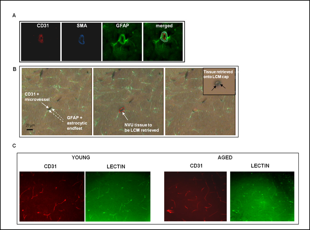
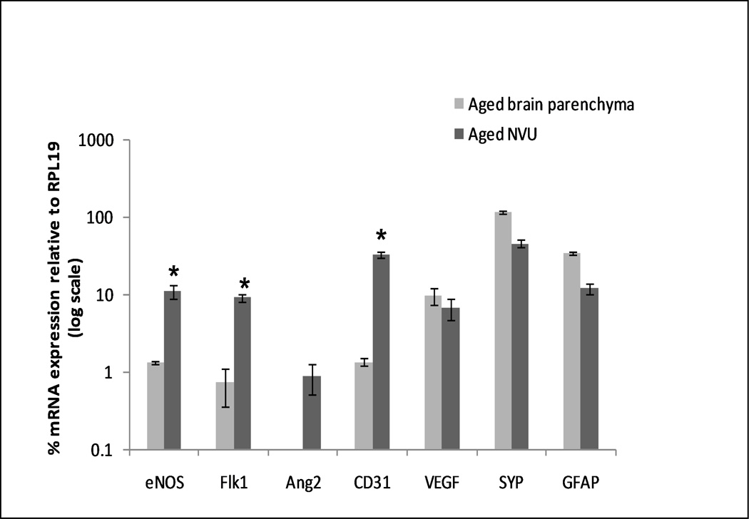
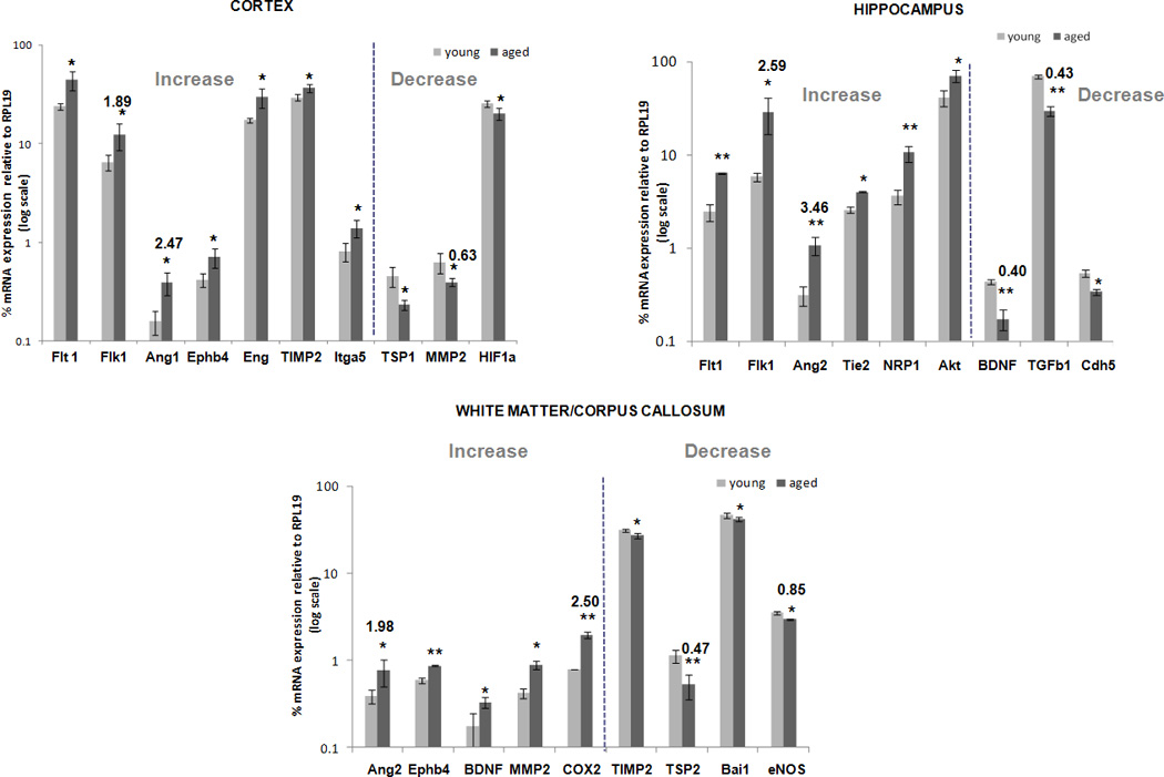
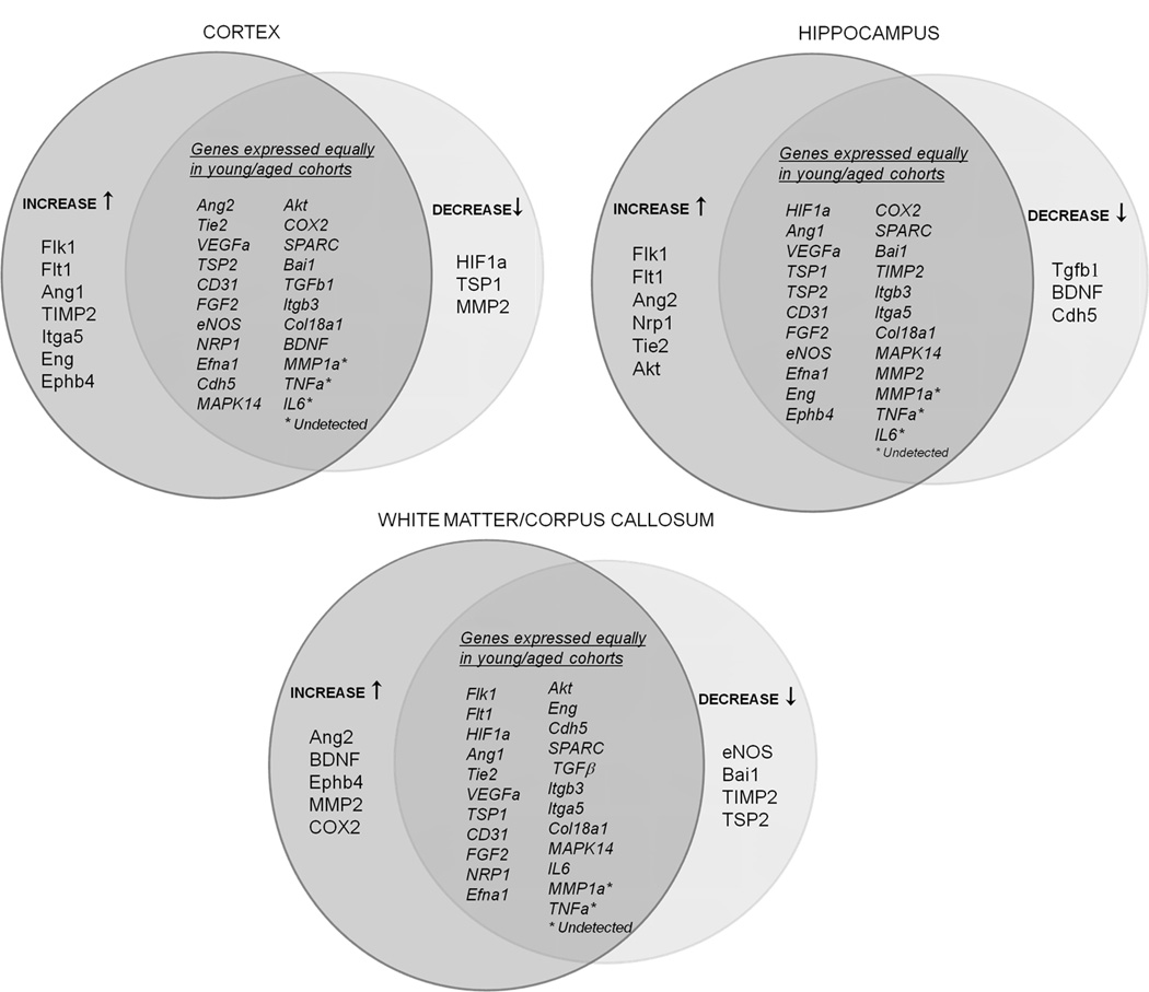
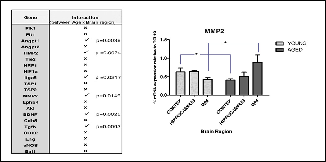
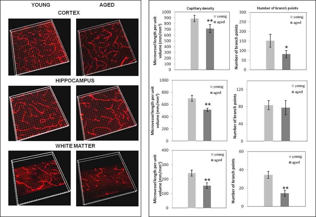
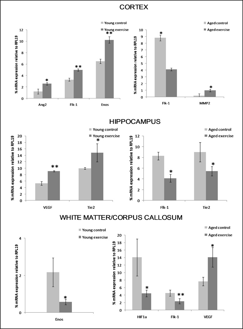
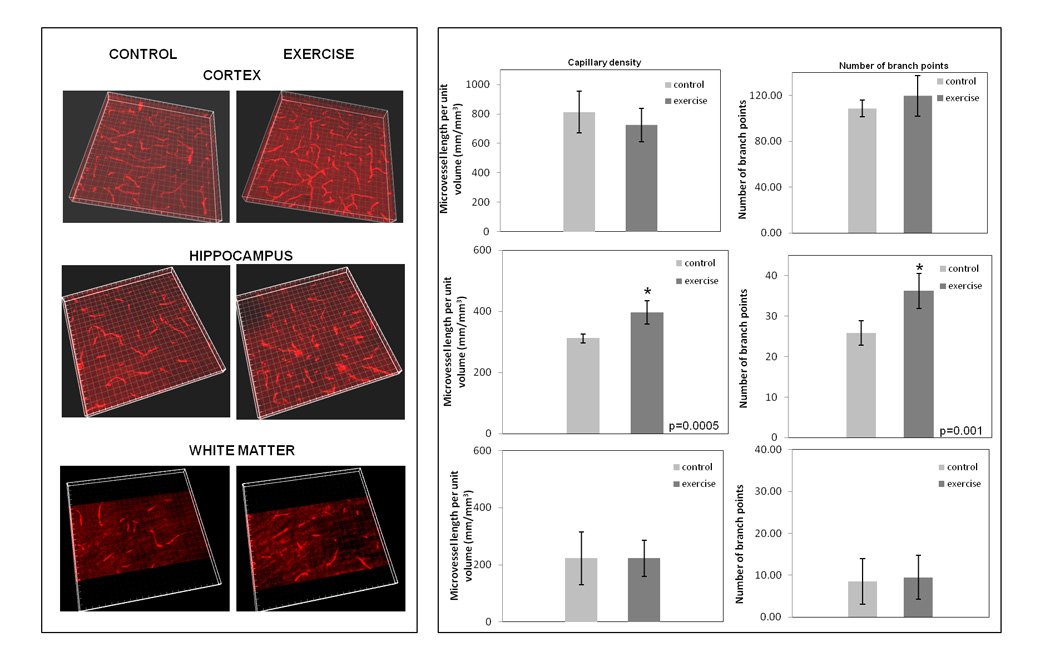
Given strong regional specialization of the brain, cerebral angiogenesis may be regionally modified during normal aging. To test this hypothesis, expression of a broad cadre of angiogenesis-associated genes was assayed at the neurovascular unit (NVU) in discrete brain regions of young versus aged mice by laser capture microdissection coupled to quantitative real-time polymerase chain reaction (PCR). Complementary quantitative capillary density/branching studies were performed as well. Effects of physical exercise were also assayed to determine if age-related trends could be reversed. Additionally, gene response to hypoxia was probed to highlight age-associated weaknesses in adapting to this angiogenic stress. Aging impacted resting expression of angiogenesis-associated genes at the NVU in a region-dependent manner. Physical exercise reversed some of these age-associated gene trends, as well as positively influenced cerebral capillary density/branching in a region-dependent way. Lastly, hypoxia revealed a weaker angiogenic response in aged brain. These results suggest heterogeneous changes in angiogenic capacity of the brain during normal aging, and imply a therapeutic benefit of physical exercise that acts at the level of the NVU.


Conflict of interest statement

Disclosure statement

The authors have no actual or potential conflicts of interest. All animal treatments were reviewed and approved by the Institutional Animal Care and Use Committee and were in accordance with guidelines stipulated by the Animal Care and Use Guidelines of the University of Connecticut Health Center and the Yale University Animal Care Committee.

Figures

Figure 1
Figure 1. The NVU can be targeted by immuno-LCM
A, Components of the NVU. A coronal section of cortex was triple-stained with antibodies to CD31 to highlight brain microvascular endothelial cells, α-SMA (alpha smooth muscle actin) to mark pericytes/smooth muscle cells, and GFAP (glial fibrillary acidic protein) to identify astrocyte foot-processes). Neuronal processes, not indicated here, are enmeshed with those of astrocytes. The - - - circle (white) encloses the NVU, the target material captured using immuno-LCM. B, Capture of NVU tissue. Left panel, Section showing CD31+ (dark brown) microvessel and surrounding GFAP+ (green) astrocyte foot-processes. Central panel, Same section with LCM “shot” over the NVU tissue (outlined in red). Right panel, Section post-LCM, showing the missing microvessel that was captured, and parenchyma left intact. Inset of right panel, Captured microvessel with closely apposed astrocyte foot-processes; i.e., NVU, on the LCM cap. C, Cerebral microvascular patency is similar in young vs. aged brain. Following labeling of patent vessels with i.v. injected FITC-lectin (green), young/aged brain cortical sections were immunostained for CD31 (red) to mark all endothelial cells. Quantitative analysis of co-localization of lectin-stained (green) and CD31+ (red) vessels was performed using the ImageJ 1.44 co-localization analysis (intensity correlation) plugin and revealed no age-related change in vessel patency.
Figure 2
Figure 2. Immuno-LCM/qRT-PCR of NVU reveals enriched expression of angiogenesis-associated genes
The graph contrasts gene expression in brain parenchymal vs. NVU tissue. Samples of either parenchymal (random LCM shots) or “targeted” NVU tissue were obtained by immuno-LCM from aged brain cortex, and processed for RNA profiling by qRT-PCR. Expression of genes is plotted as % relative to the housekeeping gene RPL19 ± S.E. (in log scale). Angiogenic genes show higher expression levels in the NVU. *p < 0.01, students two-tailed t-test.
Figure 3
Figure 3. Changes in brain regional expression of angiogenesis-associated genes in young vs. aged mice
Relative mRNA expression values of the thirty-two angiogenesis-associated genes listed in Suppl. Table 1 were determined in NVU tissue from young and aged brain regions using immuno-LCM/qRT-PCR. Only those genes that differed significantly between both age groups, in cortex, hippocampus, and white matter/corpus callosum, are depicted. RNA values are presented as mean percent expression relative to RPL19 (+ SEM) in log scale. * p < 0.05, ** p < 0.005, students t-test.
Figure 4
Figure 4. Relationships between age and brain region in expression of angiogenesis-associated genes
A, Venn diagrams displaying relative expression patterns of angiogenesis-associated genes in different regions of young vs. aged brain. Separated, are those genes that decreased (↓) or increased (↑) with aging; genes expressed equally in young and aged cohorts are depicted in the intersection. B, Interactive effects of age and brain region were determined by two-way ANOVA on those angiogenesis-associated genes that showed a significant difference with age in at least any one brain region (21 genes). Six genes exhibited positive interaction between age and brain region and are denoted by (✓); the remaining fifteen genes showed no significant interaction, and are labeled by (✗). Matrix metalloproteinase A (MMP2) expression pattern across brain regions in young versus aged mice NVU, is graphed, * p < 0.05.
Figure 5
Figure 5. Changes in brain regional capillary density and branching in young vs. aged mice
Sections (50 µm) of cortex, hippocampus and white matter/corpus-callosum were stained with anti-CD31 antibody and, following confocal microscopy and volume rendering of z stacks (left panel), capillary density (capillary length/unit volume) and branching were assessed. * p < 0.05 and ** p < 0.005.
Figure 6
Figure 6. Effect of physical exercise on expression of brain regional angiogenesis-associated genes in young vs. aged mice
Mice were subject to a treadmill regimen, and thereafter immuno-LCM/qRT-PCR analysis was performed to gauge regional NVU expression of a subset of angiogenesis-associated genes (from Suppl. Table 1) known to respond to physical exercise. Control (unexercised) groups were used for each age group. Depicted are only those genes in young and aged cohorts that were influenced significantly by exercise (compared to unexercised controls). * p < 0.05 and ** p < 0.005.
Figure 7
Figure 7. Effects of physical exercise on regional brain capillary density and branching in aged mice
Exercised mice underwent forced treadmill running for 30 min/day, for four weeks. Thereafter, brain sections (50 µm thickness) of cortex, hippocampus and white matter/corpus-callosum were stained with anti-CD31 antibody and capillary density and branching assessed as in Figure 5. Three-dimensional volume rendering of z stacks (left panel), and microvessel density and branching (right panel).

References

    1. Aird WC. Endothelium in health and disease. Pharmacol Rep. 2008;60(1):139–143. - PubMed
    1. Anderson BJ, Greenwood SJ, McCloskey D. Exercise as an intervention for the age-related decline in neural metabolic support. Front Aging Neurosci. 2010 Aug 13;2:pii: 30. 2010. - PMC - PubMed
    1. Arai K, Jin G, Navaratna D, Lo EH. Brain angiogenesis in developmental and pathological processes: neurovascular injury and angiogenic recovery after stroke. Febs J. 2009;276(17):4644–4652. - PMC - PubMed
    1. Aranha AM, Zhang Z, Neiva KG, Costa CA, Hebling J, Nor JE. Hypoxia enhances the angiogenic potential of human dental pulp cells. J Endod. 2010;36(10):1633–1637. - PubMed
    1. Benderro GF, Lamanna JC. Hypoxia-induced angiogenesis is delayed in aging mouse brain. Brain Res. 2011 - PMC - PubMed

Publication types

MeSH terms