. 2011 Nov;121(11):4537-47.
doi: 10.1172/JCI57850. Epub 2011 Oct 24.

The maternal immune response to fetal platelet GPIbα causes frequent miscarriage in mice that can be prevented by intravenous IgG and anti-FcRn therapies

Affiliations

The maternal immune response to fetal platelet GPIbα causes frequent miscarriage in mice that can be prevented by intravenous IgG and anti-FcRn therapies

Conglei Li et al. J Clin Invest. 2011 Nov.

Abstract

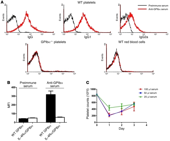
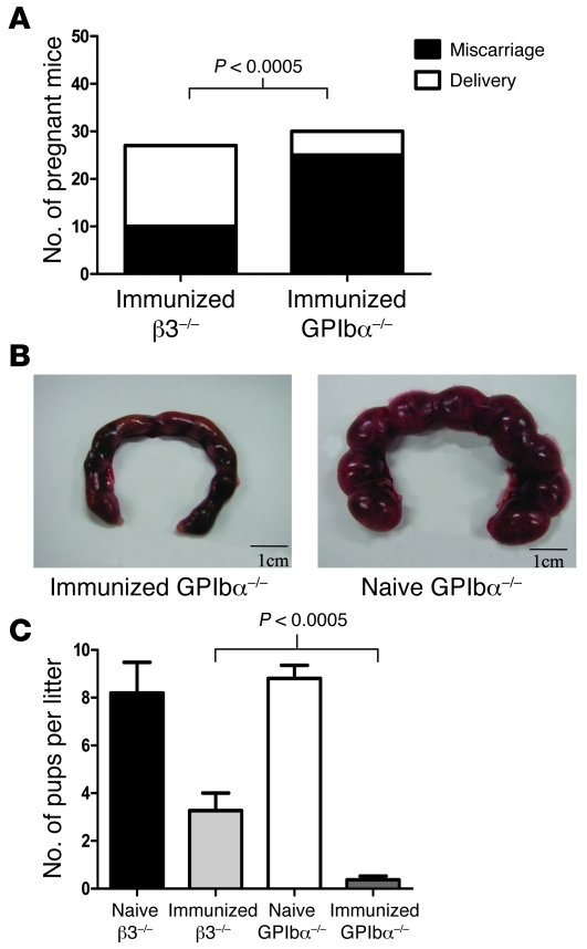
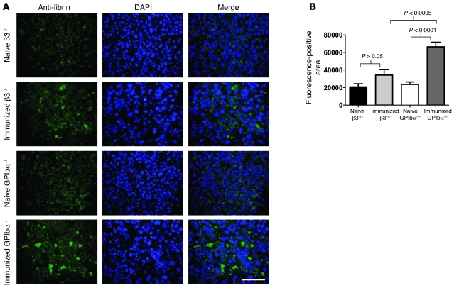
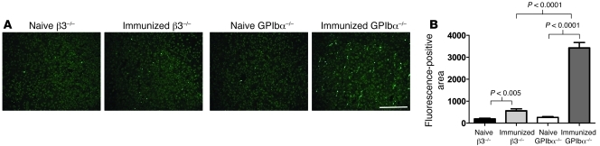
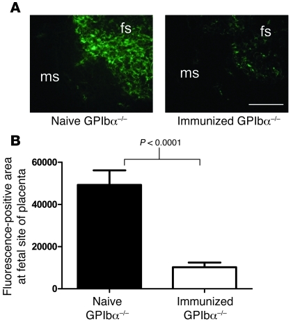
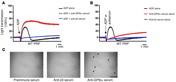
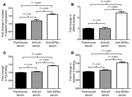
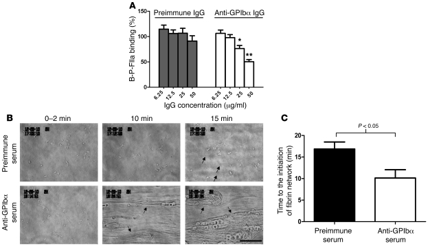
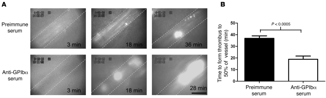
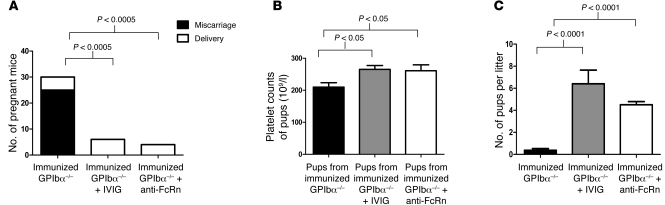
Fetal and neonatal immune thrombocytopenia (FNIT) is a severe bleeding disorder caused by maternal antibody-mediated destruction of fetal/neonatal platelets. It is the most common cause of severe thrombocytopenia in neonates, but the frequency of FNIT-related miscarriage is unknown, and the mechanism(s) underlying fetal mortality have not been explored. Furthermore, although platelet αIIbβ3 integrin and GPIbα are the major antibody targets in immune thrombocytopenia, the reported incidence of anti-GPIbα-mediated FNIT is rare. Here, we developed mouse models of FNIT mediated by antibodies specific for GPIbα and β3 integrin and compared their pathogenesis. We found, unexpectedly, that miscarriage occurred in the majority of pregnancies in our model of anti-GPIbα-mediated FNIT, which was far more frequent than in anti-β3-mediated FNIT. Dams with anti-GPIbα antibodies exhibited extensive fibrin deposition and apoptosis/necrosis in their placentas, which severely impaired placental function. Furthermore, anti-GPIbα (but not anti-β3) antiserum activated platelets and enhanced fibrin formation in vitro and thrombus formation in vivo. Importantly, treatment with either intravenous IgG or a monoclonal antibody specific for the neonatal Fc receptor efficiently prevented anti-GPIbα-mediated FNIT. Thus, the maternal immune response to fetal GPIbα causes what we believe to be a previously unidentified, nonclassical FNIT (i.e., spontaneous miscarriage but not neonatal bleeding) in mice. These results suggest that a similar pathology may have masked the severity and frequency of human anti-GPIbα-mediated FNIT, but also point to possible therapeutic interventions.


Figures

Figure 1
Figure 1. Anti-GPIbα antibodies were generated in GPIbα–/– mice after WT platelet transfusions.
(A) WT mouse platelets were incubated with a 1:100 dilution of either antiserum from GPIbα–/– mice immunized twice with 108 WT platelets or preimmune serum and stained with FITC-conjugated anti-mouse IgG, IgG1, or IgG2a. GPIbα–/– platelets and WT red blood cells were used as negative controls. (B) Anti-GPIbα serum (diluted at 1:250) specifically recognized murine platelets expressing WT GPIbα, but not IL-4Rα/GPIbα, while preimmune serum failed to recognize either. (C) Anti-GPIbα antiserum (100 μl, 50 μl, or 25 μl) was intravenously injected into WT mice, and thrombocytopenia was efficiently induced. Results are representative of 3 independent experiments (n = 3–4 mice in each group).
Figure 2
Figure 2. Miscarriage occurred in most mice of the anti-GPIbα–mediated FNIT model.
(A) GPIbα–/– and β3–/– mice were transfused twice with 108 gel-filtered WT platelets. After breeding of immunized females with WT males, far higher rates of miscarriage occurred in the anti-GPIbα–mediated FNIT model compared with the anti-β3 model (25 of 30, 83.3% versus 10 of 27, 37%; P < 0.0005). (B) The immunized GPIbα–/– mice at the time of miscarriage (around 16.5–18.5 d.p.c.) and naive GPIbα–/– mice at 17.5 d.p.c. were dissected. Much smaller conceptuses were found in the immunized GPIbα–/– females undergoing miscarriage. Scale bars: 1 cm. (C) The average number of pups per litter of females in the immunized GPIbα–/– group was smaller than that of the immunized β3–/– group (P < 0.0005).
Figure 3
Figure 3. Significantly more fibrin deposition was observed in placentas from immunized GPIbα–/– mice.
(A) Representative pictures of 14.5-d.p.c. placenta from different pregnant females (as indicated), stained with anti-fibrin antibody. Scale bar: 100 μm. (B) Quantitative analysis detected significantly more fibrin deposition in placentas from immunized GPIbα–/– mice, compared with immunized β3–/– mice (n = 6–10 mice per group).
Figure 4
Figure 4. Significantly more apoptosis/necrosis was observed in placentas from immunized GPIbα–/– mice.
(A) Representative pictures of the 14.5-d.p.c. placentas from different pregnant females with TUNEL staining. Scale bar: 200 μm. (B) Quantitative analysis revealed that significantly more apoptosis/necrosis occurred in placentas from immunized GPIbα–/– mice compared with naive GPIbα–/– mice or immunized β3–/– mice (n = 4–6 mice per group).
Figure 5
Figure 5. Significantly lower fetal blood supply was observed in placentas from immunized GPIbα–/– mice.
(A) FITC-dextran was infused into 15.5-d.p.c. pregnant GPIbα–/– mice. Representative pictures of placentas are shown. ms, maternal side; fs, fetal side. Scale bar: 200 μm. (B) Quantitative analysis of the fluorescence-positive area at the fetal side of the placenta suggested that the fetal blood supply was severely impaired in immunized pregnant GPIbα–/– mice compared with naive GPIbα–/– mice (P < 0.0001, n = 4–6 mice per group).
Figure 6
Figure 6. Anti-GPIbα antiserum cause antigen-positive platelet activation in vitro.
(A) Polyclonal anti-β3, anti-GPIbα, or preimmune serum was incubated with WT PRP prior to induction of platelet aggregation with 2 μM ADP. Anti-GPIbα serum enhanced ADP-induced WT platelet aggregation, while anti-β3 serum inhibited aggregation. (B) Anti-β3 serum alone (blue curve), anti-GPIbα serum alone (red), or ADP (2 μM; black) was added to WT PRP at 2 minutes of reaction (arrow). Anti-GPIbα, but not anti-β3, serum was able to induce mild platelet aggregation in WT PRP in the absence of soluble agonist. (C) The platelets represented in B were visualized under light microscopy. Arrows indicate platelet microaggregates. Scale bar: 50 μm. Results in AC are representative of 3 independent experiments.
Figure 7
Figure 7. Anti-GPIbα polyclonal serum induces WT platelet P-selectin expression and enhance JON/A binding and fibrinogen binding.
(AC) Polyclonal anti-β3, anti-GPIbα, or preimmune serum was incubated with the gel-filtered WT platelets and then stained by FITC-labeled anti–P-selectin antibody, PE-labeled JON/A antibody (specifically recognizing the active form of β3 integrin), or Alexa Fluor 488–labeled fibrinogen (Fg). Compared with anti-β3 serum or preimmune serum, anti-GPIbα polyclonal serum–treated WT platelets exhibited significantly more P-selectin expression, JON/A binding, and Fg binding (P < 0.0001) (n = 3–4 mice per group). (D) Anti-GPIbα polyclonal serum caused significantly more PS exposure than anti-β3 serum or preimmune serum (P < 0.0001) (n = 3–4 mice per group).
Figure 8
Figure 8. Anti-GPIbα antibodies inhibit α-thrombin binding to WT platelets, and anti-GPIbα antiserum enhances fibrin formation in vitro.
(A) The purified anti-GPIbα polyclonal antibodies significantly inhibited the binding of α-thrombin tagged with biotin/PPACK (B-P-FIIa) to murine WT platelets (*P < 0.05; **P < 0.005; P values represent comparisons between IgG and anti-GpIbα IgG groups at the same IgG concentrations, respectively). (B) Representative pictures of in vitro fibrin formation. Arrows indicate fibrin fiber formation on platelets. Scale bar: 50 μm. (C) Quantitative analysis of fibrin formation showed that anti-GPIbα polyclonal serum significantly shortened the time to form a fibrin network, compared with preimmune serum (n = 8–10 per group).
Figure 9
Figure 9. Anti-GPIbα antiserum accelerates thrombosis in vivo.
(A) Representative pictures of thrombus formation in the mesenteric artery. Images of thrombus formation were recorded for 40 minutes using a video cassette recorder. Dashed lines denote the vessel wall. Scale bar: 100 μm. (B) Quantitative analysis of thrombus formation showed that anti-GPIbα polyclonal serum significantly accelerated thrombus formation in the WT BALB/c mice compared with preimmune serum (n = 6–8 mice per group).
Figure 10
Figure 10. IVIG and anti-FcRn markedly prevented anti-GPIbα–mediated FNIT.
(A) Miscarriage in anti-GPIbα–mediated FNIT was prevented by either IVIG (0 of 5, 0% versus 25 of 30, 83.3%; P < 0.0005) or anti-FcRn treatment (0 of 4, 0% versus 25 of 30, 83.3%; P < 0.0005) when compared with untreated immunized mice. (B and C) Treatment of immunized pregnant GPIbα–/– mice with either IVIG or anti-FcRn significantly ameliorated neonatal thrombocytopenia and increased the average number of pups per litter, compared with nontreated groups.

Comment in

References

    1. Bussel JB, Primiani A. Fetal and neonatal alloimmune thrombocytopenia: progress and ongoing debates. Blood Rev. 2008;22(1):33–52. doi: 10.1016/j.blre.2007.09.002. - DOI - PubMed
    1. Kaplan C. Foetal and neonatal alloimmune thrombocytopaenia. Orphanet J Rare Dis. 2006;1:39. doi: 10.1186/1750-1172-1-39. - DOI - PMC - PubMed
    1. Blanchette VS, Chen L, de Friedberg ZS, Hogan VA, Trudel E, Decary F. Alloimmunization to the PlA1 platelet antigen: results of a prospective study. Br J Haematol. 1990;74(2):209–215. doi: 10.1111/j.1365-2141.1990.tb02567.x. - DOI - PubMed
    1. Dreyfus M, Kaplan C, Verdy E, Schlegel N, Durand-Zaleski I, Tchernia G. Frequency of immune thrombocytopenia in newborns: a prospective study. Immune Thrombocytopenia Working Group. Blood. 1997;89(12):4402–4406. - PubMed
    1. Herman JH, Jumbelic MI, Ancona RJ, Kickler TS. In utero cerebral hemorrhage in alloimmune thrombocytopenia. Am J Pediatr Hematol Oncol. 1986;8(4):312–317. doi: 10.1097/00043426-198624000-00008. - DOI - PubMed

Publication types

MeSH terms