. 2012 Jan;40(1):22-34.
doi: 10.1016/j.exphem.2011.10.003. Epub 2011 Oct 20.

A46, a benzothiophene-derived compound, suppresses Jak2-mediated pathologic cell growth

Affiliations

A46, a benzothiophene-derived compound, suppresses Jak2-mediated pathologic cell growth

Anurima Majumder et al. Exp Hematol. 2012 Jan.

Abstract

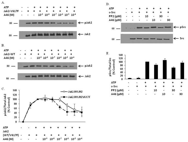
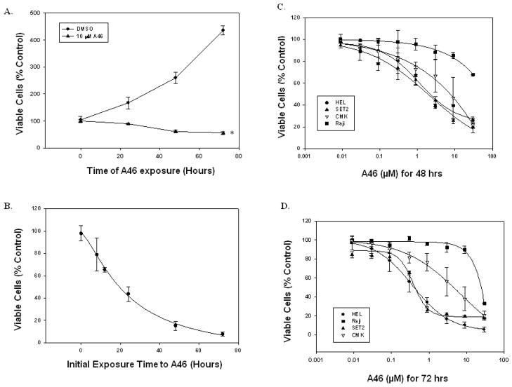
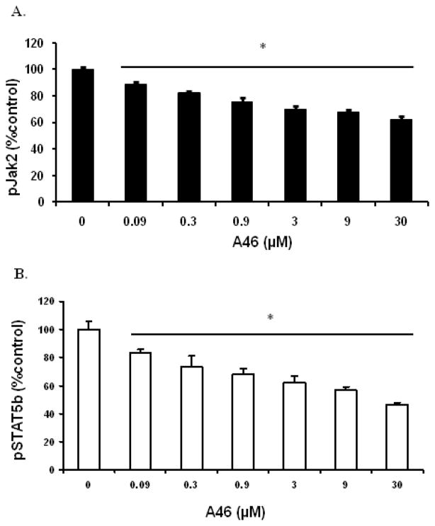
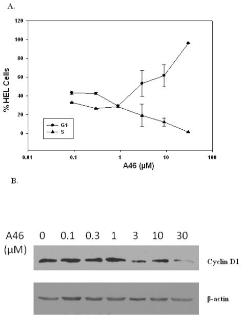
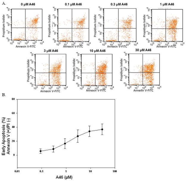
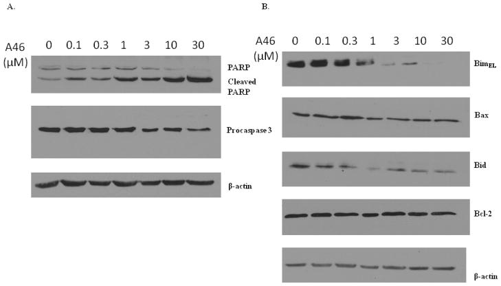
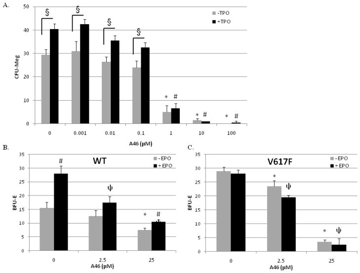
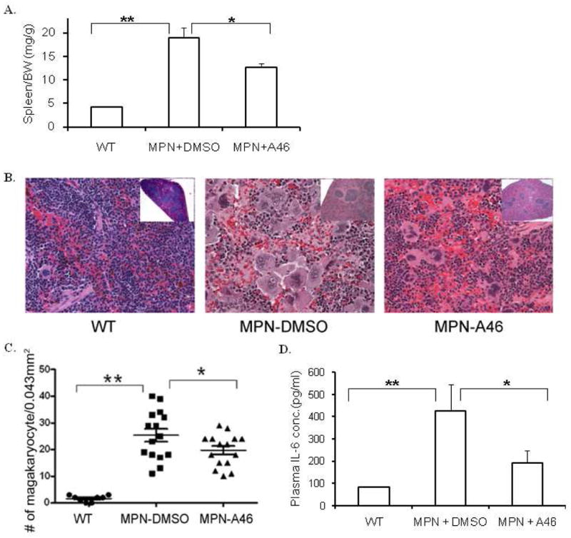
Hyperkinetic Jak2 tyrosine kinase signaling has been implicated in several hematological disorders, including myeloproliferative neoplasms. Effective Jak2 inhibitors can have significant therapeutic potential. Here, using structure-based virtual screening, we identified a benzothiophene-derived Jak2 inhibitor named A46. We hypothesized that this compound would inhibit Jak2-V617F-mediated pathologic cell growth. To test this, A46 was analyzed for its ability to inhibit recombinant Jak2 protein catalysis; suppress Jak2-mediated pathogenic cell growth in vitro; inhibit the aberrant ex vivo growth of Jak2-V617F-expressing primary human bone marrow cells; and inhibit Jak2-mediated pathogenesis in vivo. To this end, we found that A46 selectively inhibited Jak2-V617F protein when compared to wild-type Jak2 protein. The drug also selectively inhibited the proliferation of Jak2-V617F-expressing cells in both a time- and dose-dependent manner, and this correlated with decreased Jak2 and signal transducers and activators of transcription 5 phosphorylation within treated cells. The Jak2-V617F cell growth inhibition correlated with an induction of cell cycle arrest and promotion of apoptosis. A46 also inhibited the pathologic growth of primary Jak2-V617F-expressing bone marrow cells ex vivo. Lastly, using a mouse model of Jak2-V617F-mediated myeloproliferative neoplasia. A46 significantly reduced the splenomegaly and megakaryocytic hyperplasia in the spleens of treated mice and the levels of interleukin-6 in the plasma. Collectively, our data demonstrate that the benzothiophene-based compound, A46, suppresses Jak2-mediated pathogenesis, thereby making it a potential candidate drug against Jak2-mediated disorders.


Conflict of interest statement

Conflict of interest disclosure: None.

Figures

Figure 1
Figure 1. A46 selectively inhibits Jak2-V617F protein in a cell-free system
Recombinant Jak2-JH1+JH2-V617F (mutant) (A) and Jak2-JH1+JH2 (wild-type) (B) proteins were incubated in a kinase reaction buffer, either with DMSO or with the indicated doses of A46. The kinase reactions were then separated on an SDS-PAGE and immunoblotted with a phospho-Jak2 (pY1007/pY1008) antibody (A & B, top panels). The membranes were stripped and reprobed with an anti-Jak2 antibody to demonstrate equal loading of protein across all lanes (A & B, bottom panels). Shown is one of four representative results for each. C. Densitometric analysis was done on four representative Western blots to quantify A46-mediated inhibition of Jak2-V617F and Jak2 wild-type phosphorylation at the Tyr1007 residue. The ratio of phosphorylated Jak2 to total Jak2 was expressed as % control and plotted as a function of treatment condition. Shown are the means ± S.D. of four independent experiments. D. Recombinant c-Src protein was incubated in a kinase reaction buffer, either with DMSO or with the indicated doses of PP2 and A46. The kinase reactions were then separated on an SDS-PAGE and immunoblotted with a phospho-Src (pY418) antibody (D, top panel) or with an anti-Src antibody to demonstrate equal loading of protein across all lanes (D, bottom panel). Shown is one of three representative results. E. Densitometric analysis was done on the Western blots to quantify A46-mediated inhibition of c-Src phosphorylation at the Tyr418 residue. The ratio of phosphorylated Src to total Src was expressed as % control and plotted as a function of treatment condition. Shown are the means ± S.D. of three independent experiments.
Figure 2
Figure 2. A46 inhibits Jak2-V617F-dependent cell proliferation
A. HEL cells were treated with either DMSO or 10 μM A46 for 0, 24, 48 or 72 hours. Viable cell numbers for each treatment were determined by trypan blue exclusion staining using a hemocytometer. Each sample was measured in triplicate. Shown is one of two sets of representative results. *p < 0.05 with respect to DMSO. B. HEL cells were treated with 10 μM A46 for 0, 8, 12, 24, 48 and 72 hours. At the end of each treatment, the cells were washed, placed in fresh medium and cultured in the absence of the inhibitor for an additional 72 hours. The number of viable cells in each sample was then determined. Each sample was measured in triplicate. Shown are the means ± S.E. from two independent experiments. HEL, SET-2, Raji and CMK cells were treated with increasing doses of A46 for 48 (C) or 72 (D) hours. The percent viable cells from each condition were determined either by trypan blue exclusion staining. Each sample was measured in triplicate.
Figure 3
Figure 3. A46 inhibits the Jak2/STAT5 phosphorylation
HEL cells were treated with 0, 0.09, 0.3, 0.9, 3, 9 and 30 μM of A46 for 48 hours. The cell lysates were then analyzed by ELISA for the measurement of phospho-Jak2 [pY1007/pY1008] (A) and phospho-STAT5 [pY699] (B) protein levels. Each condition was run in triplicate. Shown are the means ± S.D. of two independent experiments.
Figure 4
Figure 4. A46 arrests HEL cells in G1 phase of the cell cycle
HEL cells were treated with 0, 0.1, 0.3, 1, 3, 10 and 30 μM of A46. A. After 48 hrs of treatment, cell cycle distribution of treated cells was determined as a function of drug treatment using flow cytometry. Shown are the means ± S.E. of two independent experiments. B. Following 24 hrs of treatment with the indicated concentrations of A46, cells were lysed and analyzed by immunoblotting with an anti-cyclin D1 (top) or an anti-β-actin antibody (bottom). Shown is one of two representative results.
Figure 5
Figure 5. A46 induces apoptosis in HEL cells
HEL cells were treated with 0, 0.1, 0.3, 1, 3, 10 and 30 μM of A46 for 48 hours, stained with annexin V-FITC and propidium iodide and then analyzed by flow cytometry to determine the level of apoptosis in the treated cells. A. Shown are representative flow cytometry profiles from one of two independent experiments. B. Quantification of the percentage of cells in early apoptosis as a function of drug treatment. Shown are the means ± S.E. of two independent experiments.
Figure 6
Figure 6. A46-induced apoptosis is mediated by the down regulation/cleavage of Bcl-2 family proteins Bim, Bax and Bid
HEL cells were treated with 0, 0.1, 0.3, 1, 3, 10 and 30 μM of A46 for 24 hours. The whole cell lysates were then analyzed by western blotting with a series of antibodies as indicated: A. anti-poly(ADP-ribose) polymerase (PARP), anti-caspase 3 and anti-β-actin, B. anti-Bim, -Bax, -Bid, -Bcl-2 and -β-actin. Shown are the results from one of two independent experiments for each.
Figure 7
Figure 7. A46 suppresses cytokine-independent pathologic cell growth of Jak2-V617F positive bone marrow cells, ex vivo
A. Patient-derived bone marrow mononuclear cells were cultured in semisolid medium in the presence of increasing doses of A46, with or without thrombopoietin (TPO). At the end of 14 days, the numbers of megakaryocyte colony-forming units (CFU-Megs) were counted and plotted as a function of treatment condition. Each condition was measured in duplicate. *p < 0.05 with respect to 0 μM A46 (−TPO), #p < 0.05 with respect to 0 μM A46 (+TPO), and §p < 0.05 (+ TPO) vs. (−TPO). B and C. Bone marrow aspirates obtained from either Jak2-WT or Jak2-V617F transgenic mice were treated with 0, 2.5, or 25 μM A46 for 24 hours, ex vivo. The drug was then washed away and the cells were cultured in drug-free semi-solid medium for an additional 5 days. The numbers of erythroid burst-forming units (BFU-E) in each sample were then plotted as a function of genotype. Each point was measured in duplicate. *, p<0.05 vs. − EPO 0 μM control;, p<0.05 vs. + EPO 0 μM control; #, p<0.05 vs. − EPO, within same drug treatment group.
Figure 8
Figure 8. A46 treatment alleviates Jak2-V617F mediated pathogenesis in vivo
A. Spleen weight to body weight ratios of wild-type, vehicle treated and A46 treated MPN transgenic animals. B. Representative images of H&E stained spleen histological sections from each of these different treatment groups at 100X (Inset images: 4X). C. Quantification of the number of megakaryocytes in the spleen plotted as a function of the treatment group. D. IL-6 levels in the plasma plotted as a function of the treatment group. *p<0.05, **p<0.01.

Similar articles

References

    1. Parganas E, Wang D, Stravopodis D, et al. Jak2 is essential for signaling through a variety of cytokine receptors. Cell. 1998;93:385–395. - PubMed
    1. Neubauer H, Cumano A, Muller M, Wu H, Huffstadt U, Pfeffer K. Jak2 deficiency defines an essential developmental checkpoint in definitive hematopoiesis. Cell. 1998;93:397–409. - PubMed
    1. Peeters P, Raynaud SD, Cools J, et al. Fusion of TEL, the ETS-variant gene 6 (ETV6), to the receptor-associated kinase JAK2 as a result of t(9;12) in a lymphoid and t(9;15;12) in a myeloid leukemia. Blood. 1997;90:2535–2540. - PubMed
    1. Griesinger F, Hennig H, Hillmer F, et al. A BCR-JAK2 fusion gene as the result of a t(9;22)(p24;q11.2) translocation in a patient with a clinically typical chronic myeloid leukemia. Genes Chromosomes Cancer. 2005;44:329–333. - PubMed
    1. Lacronique V, Boureux A, Valle VD, et al. A TEL-JAK2 fusion protein with constitutive kinase activity in human leukemia. Science. 1997;278:1309–1312. - PubMed

Publication types

Substances