. 2011 Nov 25;414(2):260-71.
doi: 10.1016/j.jmb.2011.10.004. Epub 2011 Oct 12.

Stepwise expansion of the bacteriophage ϕ6 procapsid: possible packaging intermediates

Affiliations

Stepwise expansion of the bacteriophage ϕ6 procapsid: possible packaging intermediates

Daniel Nemecek et al. J Mol Biol. .

Abstract

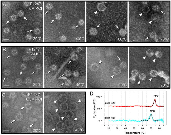
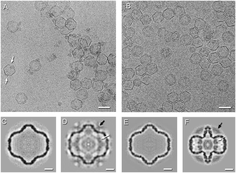
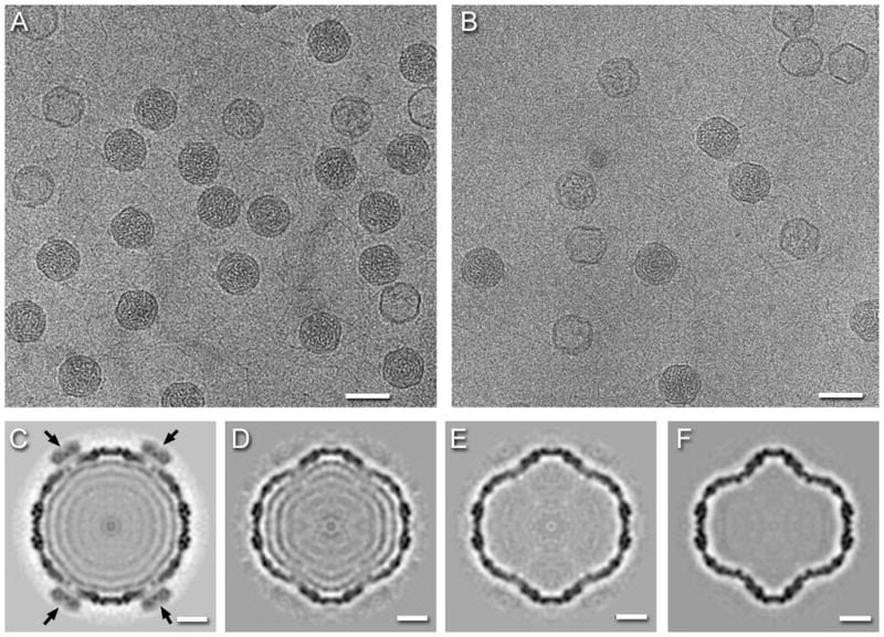
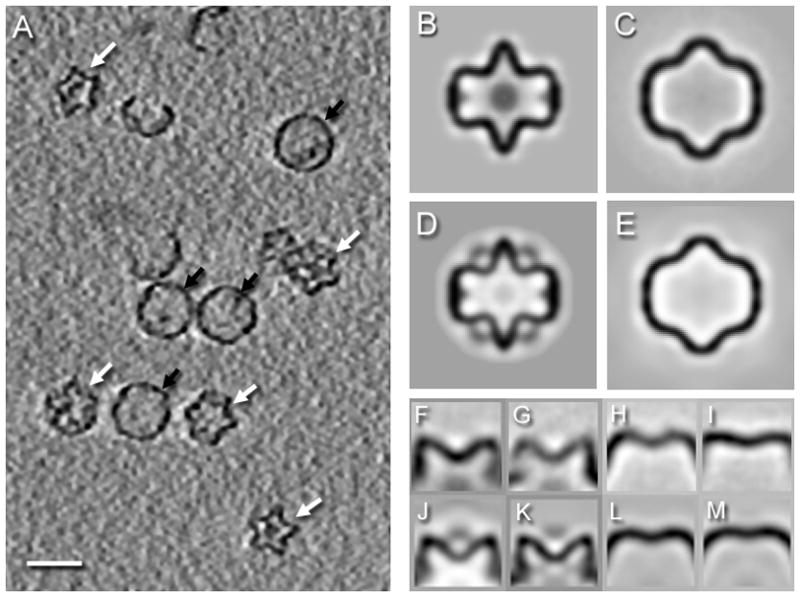
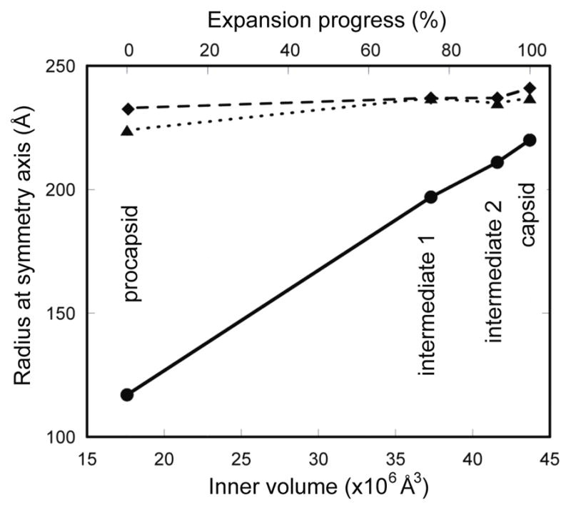
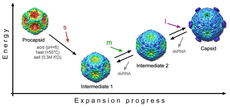
The initial assembly product of bacteriophage ϕ6, the procapsid, undergoes major structural transformation during the sequential packaging of its three segments of single-stranded RNA. The procapsid, a compact icosahedrally symmetric particle with deeply recessed vertices, expands to the spherical mature capsid, increasing the volume available to accommodate the genome by 2.5-fold. It has been proposed that expansion and packaging are linked, with each stage in expansion presenting a binding site for a particular RNA segment. To investigate procapsid transformability, we induced expansion by acidification, heating, and elevated salt concentration. Cryo-electron microscopy reconstructions after all three treatments yielded the same partially expanded particle. Analysis by cryo-electron tomography showed that all vertices of a given capsid were either in a compact or an expanded state, indicating a highly cooperative transition. To benchmark the mature capsid, we analyzed filled (in vivo packaged) capsids. When these particles were induced to release their RNA, they reverted to the same intermediate state as expanded procapsids (intermediate 1) or to a second, further expanded state (intermediate 2). This partial reversibility of expansion suggests that the mature spherical capsid conformation is obtained only when sufficient outward pressure is exerted by packaged RNA. The observation of two intermediates is consistent with the proposed three-step packaging process. The model is further supported by the observation that a mutant capable of packaging the second RNA segment without previously packaging the first segment has enhanced susceptibility for switching spontaneously from the procapsid to the first intermediate state.


Figures

Figure 1
Figure 1
Negative-stain micrographs of P1247 procapsids in (A) low-salt and (B) high-salt at different temperatures within 20–70°C as indicated. Significantly more expanded (white arrowheads) than compact (white arrows) particles were observed in the high-salt conditions. (C) Negative-stain micrographs of P1’247 procapsids in 0.3M KCl and at 20° and 40°. All particles were already expanded at 40°C and no intact particles were detected at 60°C or higher temperatures. Scale bars: 300 Å. (D) Differential scanning calorimetry of P1247 procapsids in low salt (blue curve) and 0.3 M KCl (red curve) buffers. The endothermic peaks were fitted with a non-two state model (black curves).
Figure 2
Figure 2
Cryo-electron micrographs of the P1’247 mutant procapsids at pH 5.0 (A) and of the P1247 wildtype procapsids heated to 60°C in 0.3 M KCl (B). Almost all particles appear expanded, however at the lower pH, some exhibit signs of disassembly at the 2-fold axes (white arrows). Scale bar: 500 Å. Single particle reconstructions of procapsids. (C) The spontaneously expanded P1’247 particle at pH 7.5. (D) The acid-induced expanded P1’247 particle at pH 5.5. (E) The expanded P1247 particle at 60°C and 0.3M KCl. (F) The compact P1’247 procapsid at pH 7.5. The arrows indicate densities associated with the RNA-dependent RNA polymerase, P2 (white), and packaging NTPase, P4 (black). Scale bars: 100 Å.
Figure 3
Figure 3
Cryo-electron micrographs of RNA-packaged P1247 capsids in (A) 0.1 M NaCl, and (B) 0.2 M NaCl. Scale bars: 500 Å. Reconstructions of the capsid in (C) 0.1 M NaCl and (D–E) three forms in 0.2 M NaCl showing partial or complete RNA-release. The black arrows indicate high densities of P4 subunits in the packaged capsid (C). Scale bars: 100 Å.
Figure 4
Figure 4
(A) Section through a denoised tomogram of P1247 procapsids at 60 °C in 0.3 M KCl, showing both compact (white arrows) and expanded (black arrows) particles. Scale bar: 500 Å. Central sections of icosahedral maps of the compact procapsid (B) and the expanded particle (C) of P1247 procapsids averaged from 120 and 174 non-denoised subtomograms, and with resolution estimates of 44 Å and 46 Å, respectively (FSC0.3). As a comparison, central sections through suitably resolution-limited single particle reconstructions of (D) the compact procapsid and (E) the first expansion intermediate are shown. Side views of 5-fold averaged vertex classes from subtomograms of compact procapsids (F,G) and expanded particles at 60°C (H,I). For comparison, average vertices from a single particle reconstruction (J) and a subtomogram average (K) of a compact P1247 procapsid at 22°C{Nemecek, 2010 #12573} and average vertices from single particle reconstructions of expansion intermediates 1 (K) and 2 (L) obtained from RNA-packaged procapsids after release of RNA.
Figure 5
Figure 5
(A) The procapsid expansion is largely an outward movement of the five-fold vertices, showing a linear relationship with the internal volume. The radius was measured at the three symmetry axes: 5-fold (solid line), 3-fold (dotted line), 2-fold (dashed line). Procapsid expansion is also shown in Supplementary movies 1 and 2.
Figure 6
Figure 6
Sequential steps of procapsid expansion. Here we tentatively correlate the structures produced by perturbing the procapsid or the RNA-filled capsid in vitro by environmental factors (acid, heat and/or salt) with packaging intermediates that it has not yet been possible to observe directly. The procapsid transforms in a cooperative manner to the intermediate 1 state in vitro in response to any of several treatments or, we hypothesize, by packaging the s segment. Further transformation to the intermediate 2 state would then be driven by packaging the m segment. Finally, packaging of the l segment and RNA replication yield the fully expanded capsid. To date, intermediate 2 has only been observed on perturbing the mature capsid and not on driving expansion further past the intermediate 1 state. This observation suggests that the lowest free energy state of the P1 shell in the absence of packaged RNA is intermediate 1. The reconstructions are color-coded according to the radial distance from the particle center from red to blue to emphasize the main conformational change: the outward movement of the five-fold vertices. Vertex-mounted P4 hexamers are present on the capsid reconstruction and are inferred to have been shed from the particles used to calculate the intermediate 1 and 2 reconstructions (see Discussion).

References

    1. Mindich L. Packaging, replication and recombination of the segmented genome of bacteriophage Phi6 and its relatives. Virus Res. 2004;101:83–92. - PubMed
    1. McDonald SM, Patton JT. Assortment and packaging of the segmented rotavirus genome. Trends Microbiol. 2011;19:136–44. - PMC - PubMed
    1. Sun S, Rao VB, Rossmann MG. Genome packaging in viruses. Curr Opin Struct Biol. 2010;20:114–20. - PMC - PubMed
    1. Noda T, Kawaoka Y. Structure of influenza virus ribonucleoprotein complexes and their packaging into virions. Rev Med Virol. 2010;20:380–91. - PMC - PubMed
    1. Olkkonen VM, Bamford DH. The nucleocapsid of the lipid-containing double-stranded RNA bacteriophage phi 6 contains a protein skeleton consisting of a single polypeptide species. Journal of Virology. 1987;61:2362–7. - PMC - PubMed

Publication types

LinkOut - more resources