Review
. 2012 Feb;142(2):107-16.
doi: 10.1016/j.clim.2011.09.011. Epub 2011 Oct 6.

Galectin-1 research in T cell immunity: past, present and future

Affiliations
Review

Galectin-1 research in T cell immunity: past, present and future

Filiberto Cedeno-Laurent et al. Clin Immunol. 2012 Feb.

Abstract

Galectin-1 (Gal-1) is one of 15 evolutionarily conserved ß-galactoside-binding proteins that display biologically-diverse activities in pathogenesis of inflammation and cancer. Gal-1 is variably expressed on immune cells and endothelial cells, though is commonly found and secreted at high levels in cancer cells. It induces apoptosis in effector T cells through homodimeric binding of N-acetyllactosamines on membrane glycoproteins (Gal-1 ligands). There is also compelling evidence in models of cancer and autoimmunity that recombinant Gal-1 (rGal-1) can potentiate immunoregulatory function of T cells. Here, we review Gal-1's structural and functional features, while analyzing potential drawbacks and technical difficulties inherent to rGal-1's nature. We also describe new Gal-1 preparations that exhibit dimeric stability and functional activity on T cells, providing renewed excitement for studying Gal-1 efficacy and/or use as anti-inflammatory therapeutics. We lastly summarize strategies targeting the Gal-1-Gal-1 ligand axis to circumvent Gal-1-driven immune escape in cancer and boost anti-tumor immunity.


Figures

Figure 1
Figure 1. Gal-1 induces the synthesis of IL-10+ T cells through direct and indirect mechanisms
(a) Gal-1 binding to poly-LacNAc glycans (blue antennae) on the surface of antigen presenting cells (APCs) triggers the release of IL-27, which in turn, binds to the IL-27 receptor (IL-27r) on T cells, favoring the synthesis of IL-10. (b) Alternatively, stably dimeric Gal-1 preparations directly induce T cells to synthesize IL-10, though the Gal-1 inducing mechanism is still unclear and under intense investigation.
Figure 2
Figure 2. Targeting the Gal-1 – Gal-1 ligand axis in tumors abrogates Gal-1-mediated immune privilege
(a) Tumor-derived Gal-1 induces apoptosis on anti-tumor immunocytes, and skews the cytokine milieu towards a Th2 profile, promoting tumor growth. (b) Novel approaches to interfere with the Gal-1–Gal-1 ligand axis include competitive inhibition with thiodigalactoside (TDG), a lactose-based polysaccharide; neutralization with anti-Gal-1 monoclonal antibodies and metabolic inhibition of Gal-1 ligands through fluorinated sugar analogs (e.g. fully acetylated 4-fluoro-glucosamine).
Figure 3
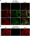
Figure 3. Gal-1-binding N-acetyllactosamines are abundantly expressed in para-cortical T cell zones in a DNFB-induced model of T cell activation
(a) Lymph nodes (LNs) draining dinitrofluorobenzene (DNFB)-sensitized skin were harvested and embedded in paraffin, and 4μm sections were stained with rabbit IgG anti-mouse CD3 (1:2000) (Abcam, San Francisco, CA) and Gal-1hFc2 (green) (20μg/ml) for 1h and counterstained with Cy-3 (red) anti-rabbit IgG (1:200) (Invitrogen, Carlsbad, CA) and rabbit anti-human IgG (1:2000) (Abcam, San Francisco, CA), followed by a 30 min incubation with Cy-5 anti-rabbit IgG (1:200) (Invitrogen, Carlsbad, CA). Note the absence of Gal-1hFc2 staining on B cell zones (dotted lines), and the clustering pattern of T cells decorated with Gal-1 ligands, suggestive of clonal expansion (inset). Representative microphotographs taken at 4X (upper and middle panels) and 10x (lower panel) are shown (Inset = 40x) (bars =100μm). (b) β-galactoside-specific Gal-1 ligand recognition on activated T cells is validated by using anti-CD3 (red) and a non-binding mutant form of Gal-1hFc2, dmGal-1hFc2 (green). Representative microphotographs from 3 experiments are shown (bars =200μm).

References

    1. Gray CA, Adelson DL, Bazer FW, Burghardt RC, Meeusen EN, Spencer TE. Discovery and characterization of an epithelial-specific galectin in the endometrium that forms crystals in the trophectoderm. Proc Natl Acad Sci U S A. 2004;101:7982–7. - PMC - PubMed
    1. Yang RY, Rabinovich GA, Liu FT. Galectins: structure, function and therapeutic potential. Expert Rev Mol Med. 2008;10:e17. - PubMed
    1. Kopcow HD, Rosetti F, Leung Y, Allan DS, Kutok JL, Strominger JL. T cell apoptosis at the maternal-fetal interface in early human pregnancy, involvement of galectin-1. Proc Natl Acad Sci U S A. 2008;105:18472–7. - PMC - PubMed
    1. Rabinovich GA, Toscano MA. Turning 'sweet' on immunity: galectin-glycan interactions in immune tolerance and inflammation. Nat Rev Immunol. 2009;9:338–52. - PubMed
    1. Camby I, Le Mercier M, Lefranc F, Kiss R. Galectin-1: a small protein with major functions. Glycobiology. 2006;16:137R–157R. - PubMed

MeSH terms