. 2011 Dec 15;360(2):358-68.
doi: 10.1016/j.ydbio.2011.10.005. Epub 2011 Oct 12.

The first bromodomain of the testis-specific double bromodomain protein Brdt is required for chromocenter organization that is modulated by genetic background

Affiliations

The first bromodomain of the testis-specific double bromodomain protein Brdt is required for chromocenter organization that is modulated by genetic background

Binyamin D Berkovits et al. Dev Biol. .

Abstract

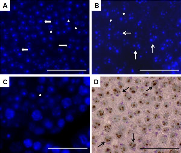
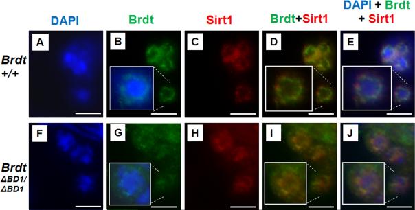
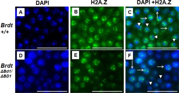
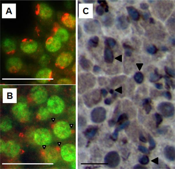
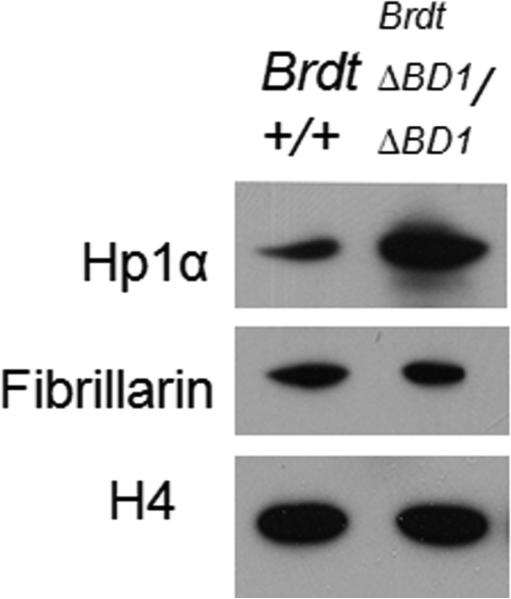
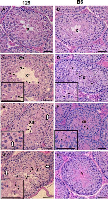
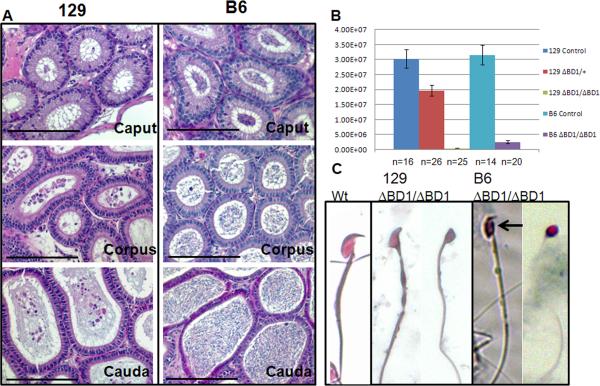
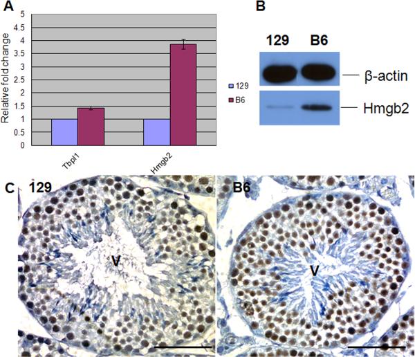
Mice homozygous for a mutation (Brdt(∆BD1/∆BD1)) lacking the first bromodomain of Brdt, a testis-specific member of the BET family of double-bromodomain containing proteins, are sterile and exhibit profound defects in chromatin remodeling during spermiogenesis. We have now observed that a prominent feature of the aberrant spermatid nuclei is a fragmented chromocenter, a structure comprised of peri-centromeric heterochromatin. There was a concomitant increase in the levels of heterochromatin protein 1 alpha (Hp1α), suggesting that the presence of multiple chromocenters was correlated with a spread of heterochromatin beyond the normal centromeric region. Brdt protein was normally present throughout the nucleus but was excluded from the chromocenter. A more densely staining region of Brdt protein appeared to separate sirtuin 1 (Sirt1) protein from contact with the chromocenter. Although still nuclear, this unique localization of Brdt protein was lost in Brdt(∆BD1/∆BD1) mutant spermatids and Brdt and Sirt1 overlapped around the chromocenters. There was also ectopic localization of the H1 histone family, member N, testis-specific (H1fnt) protein in Brdt(∆BD1/∆BD1) round spermatids, which may be linked to the previously reported loss of polarized localization of peri-nuclear heterochromatin foci. The extent of chromocenter fragmentation was more severe and penetrant in mutant testes on a pure 129Sv/Ev as compared to a pure C57Bl/6 background. Indeed, all aspects of the mutant phenotype were more severe on the 129Sv/Ev background. Contrary to previous studies in genetic models where fragmented chromocenters were observed in spermatids, the Brdt(∆BD1/∆BD1) mutant spermatids do not undergo apoptosis (on either background). These observations suggest that the first bromodomain of Brdt is critical in the formation and/or maintenance of an intact chromocenter and implicate this structure in proper remodeling of the chromatin architecture of the sperm head.


Figures

Figure 1
Figure 1. BrdtΔBD1/ΔBD1 round spermatids exhibit severe fragmentation of the chromocenter
(A-C) DAPI staining of histological sections from post-natal day 24 testes (A) 129 control, (B) mutant and (C) B6 mutant. Wide arrows: two foci of heterochromatin, normal. Arrowheads: three foci, fragmented chromocenter. Arrows: four or more foci, severely fragmented chromocenter. The number of spermatids with multiple chromocenters seen in the mutant 129 testes is greater than that seen in control or in the B6 mutant (also see Table 1). (D) Immunostaining of histone 3 trimethylated on lysine 9 and periodic acid Schiff (PAS) staining of the acrosome of day 24 mutant 129 testis. The cells with severe fragmentation also have acrosomes and thus are round spermatids (arrows). Scale bar, 50 μm.
Figure 2
Figure 2. Dense Brdt expression separates the chromocenter from Sirt1 protein
Fluorescent staining of control (A-E) and mutant (F-J) round spermatids with Brdt and Sirt1 antibodies and the merge of the two.(A-B) Brdt is present in the nucleus of round spermatids, with regions of denser staining surrounding the chromocenter from which it is excluded (inset merge of Brdt and DAPI). (C) Sirt1 is also present in the nucleus of round spermatids, and is also excluded from the chromocenter (inset in E). (D-E) Brdt and Sirt1 protein localization overlap except for the region directly surrounding the chromocenter where Brdt is strongly detected but Sirt1 is excluded (green fluorescence only). (F-G) Truncated Brdt protein is still present in the nucleus of mutant round spermatids, but the denser staining around the chromocenter is no longer seen (inset merge of Brdt and DAPI). (H) Sirt1 expression is still nuclear and excluded from the chromocenter. (I-J) Brdt and Sirt1 protein localization overlap completely (no exclusive green fluorescence), and both are present directly adjacent to the chromocenter. While there are still some regions of dense Brdt staining, it is no longer localized around the chromocenter. Scale bar, 12.5 μm.
Figure 3
Figure 3. Localization of H2A.Z around the chromocenter is unchanged in BrdtΔBD1/ΔBD1 mutant round spermatids
Fluorescent staining of DAPI (A,D), H2A.Z (B,E) and merge (C,F) in control (A-C) and mutant (D-F) day 24 histological sections. (A-C) In control spermatids H2A.Z is localized around the intact chromocenter (arrowheads). Fragmented chromocenters still have H2A.Z surrounding the foci of heterochromatin (arrows). (D-F) In mutant round spermatids H2A.Z is localized normally, both around intact chromocenters (arrowheads) and severely fragmented chromocenters (arrows). Scale bar, 50 μm.
Figure 4
Figure 4. H1fnt is ectopically localized in some 129 mutant round spermatids
(A-B) Fluorescent staining to localize H1fnt in day-24 histological sections reveals a single region of apical expression in control 129 round spermatids (A) and ectopic localization in mutant 129 round spermatids (B, open arrowheads). (C) Immunostaining of H1fnt in a mutant 129 adult histological section shows non-elongated mutant spermatids with H1fnt surrounding the nucleus (arrowheads). Scale bar, 25 μm.
Figure 5
Figure 5. There is an increase in the amount of heterochromatin in the mutant testes
Chromatin was extracted from control and mutant spermatogenic cells and immunoblotted with anti-Hp1α antibody, anti-fibrillarin antibody and anti-H4 antibody. Over 3-fold more Hp1α is present in mutant chromatin. Fibrillarin levels were unchanged in the mutant testes. H4 was used as a loading control.
Figure 6
Figure 6. The abnormalities in BrdtΔBD1/ΔBD1 mice are more severe and uniform on the 129 background
(A-H) H&E staining of histological sections of (A) control129 stage X tubule, (B) control B6 stage X tubule, (C) mutant 129 stage X* tubule, (D) mutant B6 stage X tubule, (E) mutant 129 stage XII* tubule, (F) mutant B6 stage XII tubule, (G) mutant 129 stage V* tubule, and (H) mutant B6 stage V tubule. Grossly normally elongating spermatids are absent in the mutant 129 testes, but predominate the B6 mutant testis. Conversely, compacted but not elongated spermatids are present (C-H, arrowheads and D-G inset) in B6 testes, but they predominate the 129 testes. (see Table 2 for quantification). Spermatids which have neither compacted nor elongating (C, arrow and inset) can be found only in the 129. In 129 testes some spermatids can be found in the spermatocyte layer of the tubules (C,E, and G, open arrows and E inset). Asterisk: although the severe disruption of the order of the seminiferous epithelium in the 129 mutant made precise staging difficult, we refer to approximately staged tubules with a roman numeral followed by an asterisk. Scale bar, 25 μm.
Figure 7
Figure 7. Differences in epididymal sperm content of BrdtΔBD1/ΔBD1 mutants on 129 and B6 backgrounds
(A) H&E staining of histological sections of mutant 129 and mutant B6 caput, corpus and caudal epididymides, showing the nearly complete absence of sperm in the 129 animal and the abundance of sperm in the B6 animal. (B) Caudal sperm counts from control, heterozygous and homozygous mutant 129 mice and control and homozygous mutant B6 mice. There is a statistically significant loss of sperm in the 129 heterozygous cauda compared to control and a statistically significant difference between the 129 and B6 mutant counts. (C) Light microscope photomicrographs of 129 control and BrdtΔBD1/ΔBD1 and B6 BrdtΔBD1/ΔBD1 sperm. Although all sperm are abnormal, there is greater heterogeneity in the B6 sperm, with some sperm that appear close to normal (arrow). Scale bar, 50 μm.
Figure 8
Figure 8. Hmgb2 is expressed at higher levels in the B6 testis
(A) Quantitative real-time PCR showing that Hmgb2 but not Tbpl1 transcription is higher in the B6 testis than in the 129 testis. The expression levels are shown as relative levels of the 129 expression which is set as 1. (B) Western blot of Hmgb2 confirming 4 times greater protein in the B6 testis. β-actin is used as loading control. (C) Hmgb2 expression in stage V tubules. Protein levels are higher in the B6 cells, specifically in round spermatids. Scale bar, 50 μm.

References

    1. Bannister AJ, Zegerman P, Partridge JF, Miska EA, Thomas JO, Allshire RC, Kouzarides T. Selective recognition of methylated lysine 9 on histone H3 by the HP1 chromo domain. Nature. 2001;410:120–4. - PubMed
    1. Buhler M, Gasser SM. Silent chromatin at the middle and ends: lessons from yeasts. Embo J. 2009;28:2149–61. - PMC - PubMed
    1. Catena R, Ronfani L, Sassone-Corsi P, Davidson I. Changes in intranuclear chromatin architecture induce bipolar nuclear localization of histone variant H1T2 in male haploid spermatids. Dev Biol. 2006;296:231–238. - PubMed
    1. Chua P, Roeder GS. Bdf1, a yeast chromosomal protein required for sporulation. Mol Cell Biol. 1995;15:3685–96. - PMC - PubMed
    1. Chung SS, Wang X, Roberts SS, Griffey SM, Reczek PR, Wolgemuth DJ. Oral administration of a retinoic Acid receptor antagonist reversibly inhibits spermatogenesis in mice. Endocrinology. 2011;152:2492–502. - PMC - PubMed

Publication types