. 2011 Oct 23;13(11):1353-60.
doi: 10.1038/ncb2366.

miR-34 miRNAs provide a barrier for somatic cell reprogramming

Affiliations

miR-34 miRNAs provide a barrier for somatic cell reprogramming

Yong Jin Choi et al. Nat Cell Biol. .

Abstract

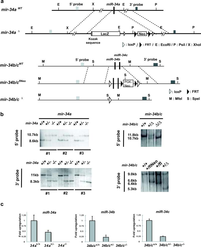
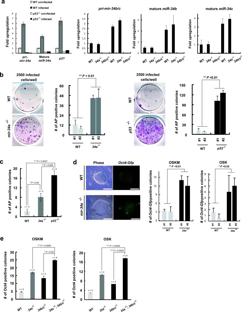
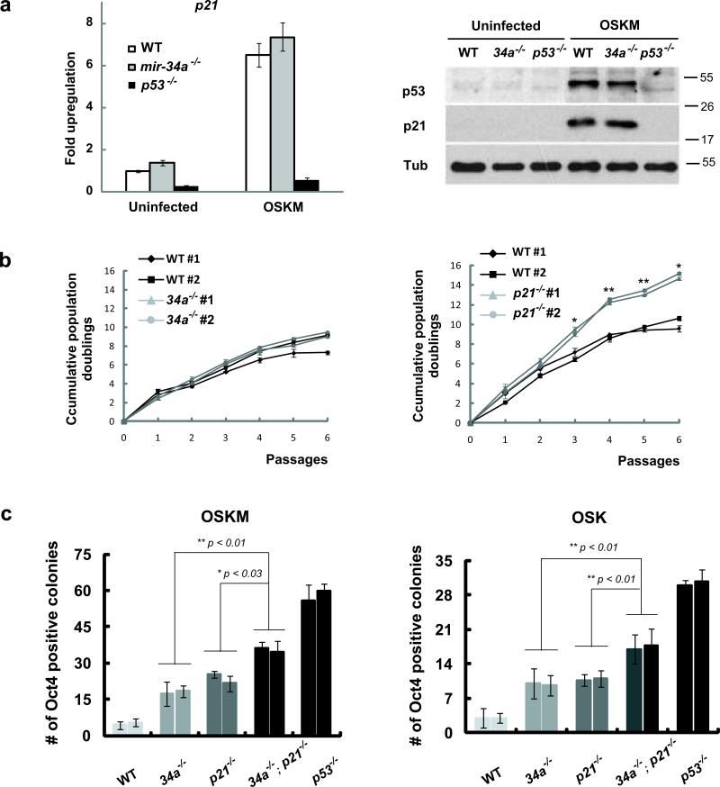
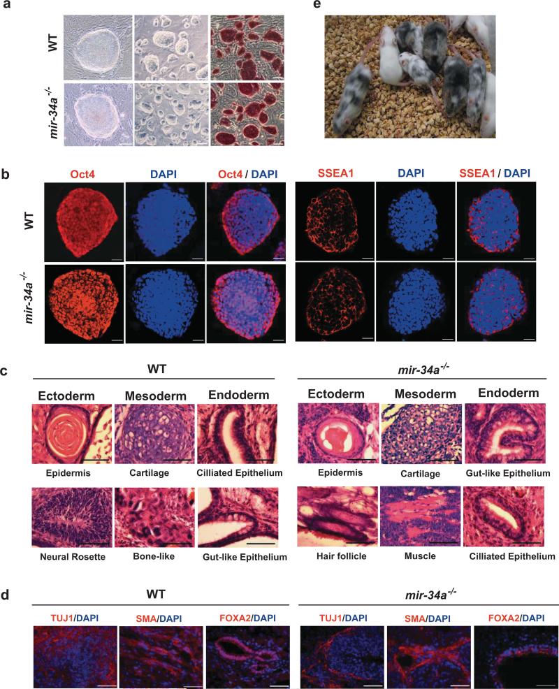
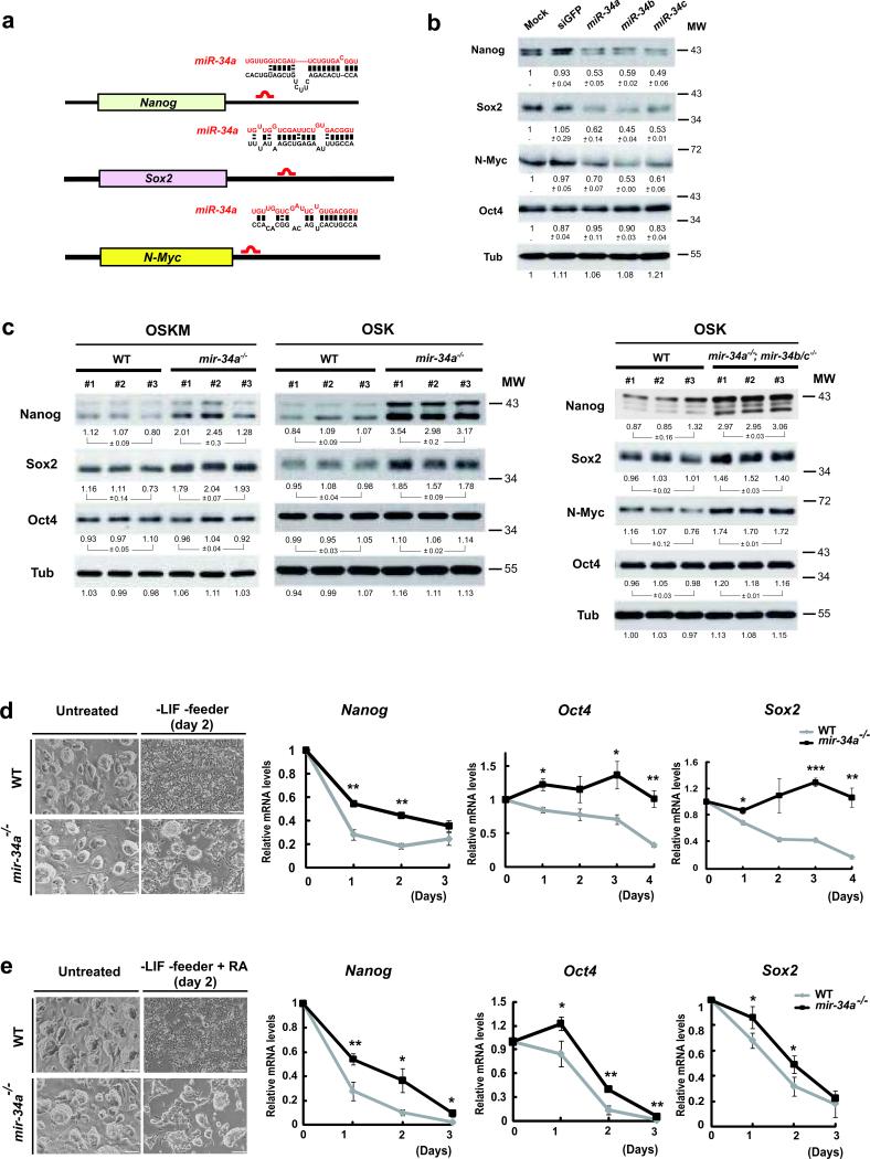
Somatic reprogramming induced by defined transcription factors is a low-efficiency process that is enhanced by p53 deficiency. So far, p21 is the only p53 target shown to contribute to p53 repression of iPSC (induced pluripotent stem cell) generation, indicating that additional p53 targets may regulate this process. Here, we demonstrate that miR-34 microRNAs (miRNAs), particularly miR-34a, exhibit p53-dependent induction during reprogramming. Mir34a deficiency in mice significantly increased reprogramming efficiency and kinetics, with miR-34a and p21 cooperatively regulating somatic reprogramming downstream of p53. Unlike p53 deficiency, which enhances reprogramming at the expense of iPSC pluripotency, genetic ablation of Mir34a promoted iPSC generation without compromising self-renewal or differentiation. Suppression of reprogramming by miR-34a was due, at least in part, to repression of pluripotency genes, including Nanog, Sox2 and Mycn (also known as N-Myc). This post-transcriptional gene repression by miR-34a also regulated iPSC differentiation kinetics. miR-34b and c similarly repressed reprogramming; and all three miR-34 miRNAs acted cooperatively in this process. Taken together, our findings identified miR-34 miRNAs as p53 targets that play an essential role in restraining somatic reprogramming.


Figures

Fig. 1
Fig. 1. Generation of mir-34a and mir-34b/c knockout MEFs
A. Diagrams of endogenous mir-34a and mir-34b/c gene structure and the knockout construct. Using recombineering, we engineered the mir-34a targeting vector with a ~6kb homologous arm on both 5’ and 3’ ends, flanking a Kozak sequence, a lacZ cDNA and a FRT-neo-FRT cassette. The mir-34b/c targeting vector contains a ~6kb homologous arm at each end, with the mir-34b/c gene and a Neo selection cassette flanked by loxP sites. b. Validating the germline transmission of the mir-34a and mir-34b/c targeted allele using Southern analysis. Putative WT, mir-34a+/- and mir-34a-/- animals derived from three independently targeted ES line were analyzed by Southern blot using probes either 5’ or 3’ to the homologous arms (left). Similar validation was performed for WT, mir-34b/c+/- and mir-34b/c-/- animals (right). c. Confirming loss of mir-34 expression in mir-34a and mir-34b/c knockout MEFs. Littermate-controlled WT, mir-34a+/- and mir-34a-/- MEFs were analyzed by real-time PCR to quantify the expression of mir-34a. While WT MEFs showed robust miR-34a induction upon culture stress, no miR-34a expression was detected in mir-34a-/- MEFs. The miR-34a level in mir-34a+/- MEFs was approximately half that of WT MEFs. Similar validation was performed for mir-34b/c-/- MEFs. Error bar, standard deviation, n=3.
Fig. 2
Fig. 2. Deficiency of miR-34 miRNAs increases reprogramming efficiency
a. Four reprogramming factors triggered p53-dependent induction of miR-34 miRNAs. Three days after transduction, pri-mir-34a, mature miR-34a and p21 were measured in uninfected and four-factor induced WT and p53-/- MEFs. Induction of pri-mir-34a was dependent on the intact p53 response, and was comparable to that of p21. Induction of pri-mir-34b/c, mature miR-34b and c was determined in WT, mir-34a-/- and mir-34b/c-/- MEFs. Error bar, standard deviation, n=3. b. mir-34a deficiency significantly enhanced three-factor induced MEF reprogramming. 2500 three-factor infected WT, mir-34a-/- or p53-/- MEFs were plated to score reprogramming by AP-positive colonies with characteristic ESC morphology. A representative image and quantitative analysis is shown out of five independent experiments, comparing littermate-controlled WT and mir-34a-/- MEFs (left, **P < 0.01), as well as WT and p53-/- MEFs (right, **P < 0.01). Error bar, standard deviation, n=4. c. Single-sorted, four-factor infected MEFs were cultured at a density of one cell per well. Four weeks post-plating, AP-positive colonies with typical iPSC morphology were scored for WT, mir-34a-/- and p53-/- iPSCs. Four independent experiments confirmed this finding. *P < 0.05 for comparison between WT and mir-34a-/- MEFs. Error bar, standard error, n= experiments with independent MEF lines. d. mir-34a deficiency significantly enhanced MEF reprogramming as measured by Oct4-Gfp reporter expression. Three- or four-factor infected WT and mir-34a-/- MEFs that carry an Oct4-Gfp allele were sorted at the density of 2500 cells/well and 1000 cells/well, respectively. Reprogramming efficiency was quantified by GFP positive clones. Images of Oct4-Gfp positive iPSCs were shown on the left. A quantitative analysis for reprogramming efficiency triggered by four factor (left, **P<0.01) or three factor (right, *P<0.02) was shown. Scale bar, 100μm. Error bar, standard deviation, n=3. OSKM, Oct4, Sox2, Klf4 and Myc; OSK, Oct4, Sox2 and Klf4. e. miR-34a, b, and c cooperatively regulate somatic reprogramming. Deficiency in mir-34a or mir-34b/c alone significantly promoted somatic reprogramming, yet deficiency in all mir-34 miRNAs exhibited further increase. Two independent experiments confirmed this finding. Error bar, standard error, n = experiments with independent MEFs. All P-values were calculated based on two-tailed Student's t-test.
Fig. 3
Fig. 3. miR-34a and p21 cooperate to repress iPSCs generation
a. p21 was induced in mir-34a-/- MEFs during somatic reprogramming. Three days after retroviral transduction of four reprogramming factors, both p21 mRNA (left) and p21 protein (right) exhibited a significant increase in WT and mir-34a-/- MEFs. This increase correlated well with the elevated level of p53 proteins (right). α-Tubulin (Tub) was used as a loading control. Error bar, standard deviation, n=3. b. p21-/- MEFs proliferate more rapidly than mir-34a-/- MEFs. Cumulative population doublings were measured for 6 consecutive passages in littermate-controlled WT and mir-34a-/- MEFs (left), and in WT and p21-/- MEFs (right). Compared to the WT counterparts, p21-/- MEFs exhibited an enhanced cell proliferation rate, while mir-34a-/- MEFs showed little differences. Error bar, standard deviation, n=3 for triplicate measurements at each time point. *P < 0.05; **P < 0.01 for comparisons between two lines of MEFs for each genotype. c. miR-34a and p21 cooperate to repress iPSCs generation. The reprogramming efficiency were compared among WT, mir-34a-/-, p21-/-, mir-34a-/-; p21-/- and p53-/- MEFs using either three (right) or four (left) reprogramming factors. Deficiency in mir-34a or p21 alone enhanced reprogramming efficiency to a comparable level. Deficiency in both mir-34a and p21 gave rise to an even greater reprogramming efficiency. Quantitative analyses of Oct4-positive colonies were carried out at 2 (four-factor induced reprogramming) or 3 (three-factor induced reprogramming) weeks post-plating using immunofluorescence analyses. Error bar, standard deviation, n=3. OSKM, Oct4, Sox2, Klf4 and Myc; OSK, Oct4, Sox2 and Klf4; MW, molecular weight. All P-values were calculated based on two-tailed Student's t-test.
Fig. 4
Fig. 4. mir-34a-/- iPSCs functionally resemble WT iPSCs
a. iPSCs derived from both WT and mir-34a-/- MEFs exhibited ES-like morphology in culture, with robust AP expression. Scale bar, 20μm for the left panel, 100 μm for the middle and right panels. b. Both WT and mir-34a-/- iPSCs expressed pluripotency markers, including nucleus-localized Oct4 and membrane-localized SSEA1. Scale bar, 20μm; c, d. Wildtype and mir-34a-/- iPSCs both generated differentiated teratomas. Teratomas derived from four-factor induced WT (left) and mir-34a-/- (right) iPSCs were harvested from nude mice 4-6 weeks after subcutaneous injection. H&E staining(c), as well as immunofluorescence staining (d), revealed terminally differentiated cell types derived from all three germ layers. Scale bar in c, 25μm; in d, 50μm. e. Four-factor-induced mir-34a-/- iPSCs efficiently contribute to adult chimeric mice. We injected three independent lines of passage seven Oct4-Gfp/+, mir-34a-/- iPSCs into albino-C57BL/6/cBrd/cBrd/cr blastocysts. The iPSC contribution to adult chimeric mice was determined by coat color pigmentation.
Fig. 5
Fig. 5. mir-34a represses Nanog, Sox2 and N-Myc expression post-transcriptionally
a. Schematic representation of the Nanog, Sox2 and N-Myc 3’UTR, and the predicted mir-34 binding sites. The mouse Nanog, Sox2 and N-Myc 3’UTR each contains one putative miR-34a binding site within their 3’UTRs. b. Enforced expression of mir-34a, b, and c in ESCs reduced the protein levels of Nanog, Sox2 and N-Myc, but not Oct4. Feeder-free ESCs were transfected with miRNA mimics for miR-34a, miR-34b and miR-34c, and a negative control, siGFP. At 48 hours post transfection, Western analysis indicated a significant reduction in the protein levels of Nanog, Sox2 and N-Myc, but not Oct4. The value of each band indicates the relative expression level normalized by the internal control, α-tubulin, averaged among two independent experiments, and presented as mean ± s.e.m. c. Derepression of Nanog, Sox2 and N-Myc was observed in mir-34 deficient iPSCs. A significant increase of Nanog and Sox2, but not Oct4, was observed in four factor induced mir-34a-/- iPSCs, when compared to passage matched, littermate controlled WT iPSCs. A similar comparison was performed for passage matched, three-factor induced WT and mir-34a -/-; mir-34b/c-/- double knockout iPSCs, where derepression of Nanog, Sox2 and N-Myc was observed. For this Western analysis, the quantitation of each band was performed by Quantity One software, and was normalized against its own internal tubulin control. The standard errors of three independent iPSC lines were shown for each genotype, n=3. d, e. mir-34a deficient iPSCs exhibited slower kinetics during differentiation. Wildtype and mir-34a-/- iPSCs were both triggered to differentiate by withdrawal of LIF in the presence (e) or absence (d) of RA treatment. The image of typical iPSC culture two days after each differentiation condition were shown on the top (d, e), and the quantitative analyses on the decline of Nanog, Sox2 and Oct4 transcripts in response to these differentiating conditions were shown on the bottom (d, e). Error bar, standard error, n=3. * P<0.05, ** P<0.01. Scale bar in d and e, 100 μm. MW, molecular weight.

References

    1. Hong H, et al. Suppression of induced pluripotent stem cell generation by the p53-p21 pathway. Nature. 2009;460:1132–1135. - PMC - PubMed
    1. Kawamura T, et al. Linking the p53 tumour suppressor pathway to somatic cell reprogramming. Nature. 2009;460:1140–1144. - PMC - PubMed
    1. Li H, et al. The Ink4/Arf locus is a barrier for iPS cell reprogramming. Nature. 2009;460:1136–1139. - PMC - PubMed
    1. Marion RM, et al. A p53-mediated DNA damage response limits reprogramming to ensure iPS cell genomic integrity. Nature. 2009;460:1149–1153. - PMC - PubMed
    1. Utikal J, et al. Immortalization eliminates a roadblock during cellular reprogramming into iPS cells. Nature. 2009;460:1145–1148. - PMC - PubMed

Publication types

MeSH terms