. 2012 Mar 1;520(4):848-73.
doi: 10.1002/cne.22793.

Development and distribution of neuronal cilia in mouse neocortex

Affiliations

Development and distribution of neuronal cilia in mouse neocortex

Jon I Arellano et al. J Comp Neurol. .

Abstract

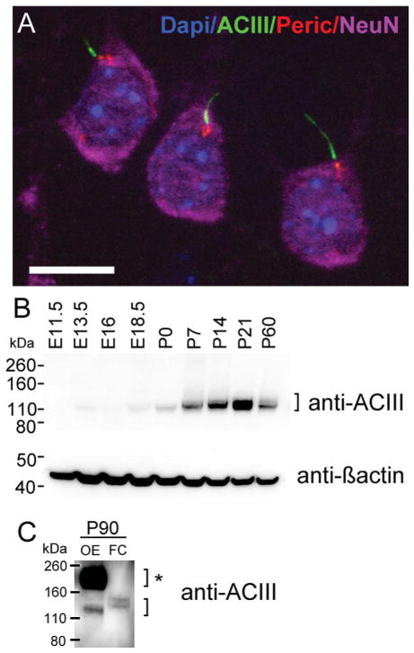
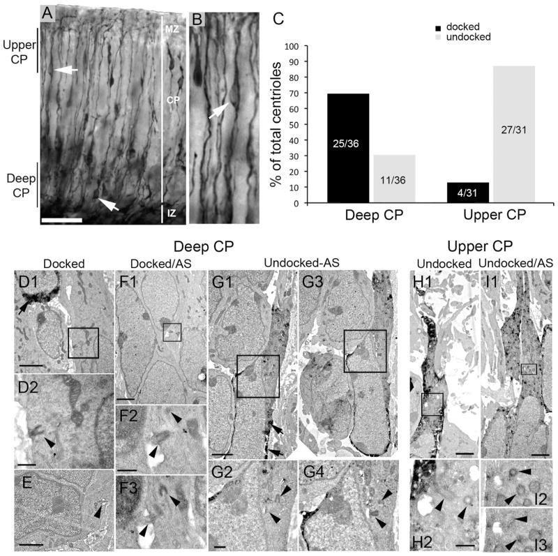
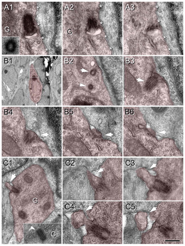
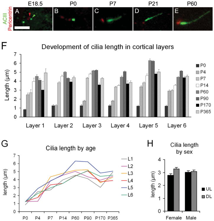
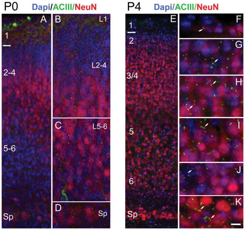
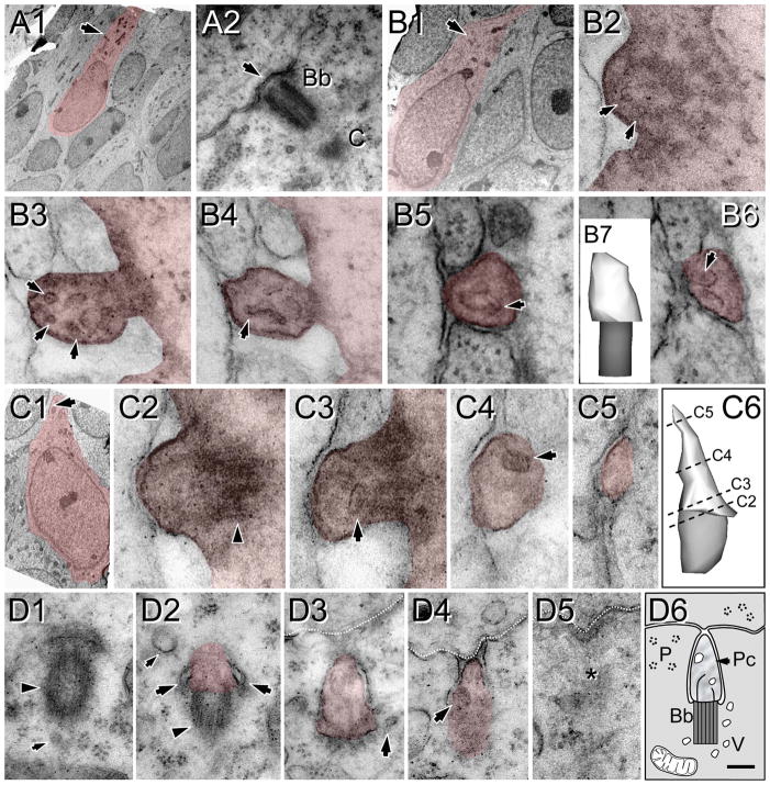
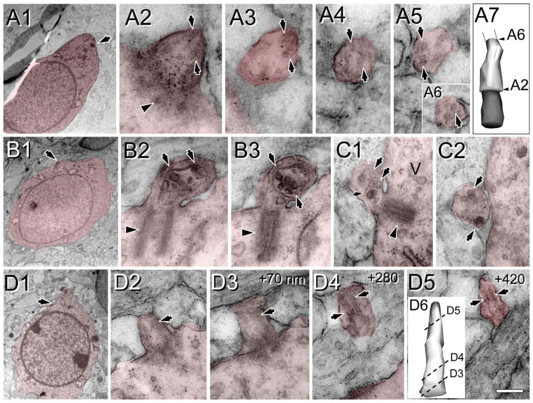
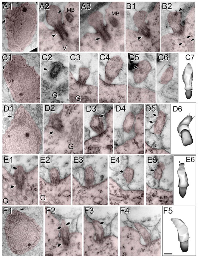
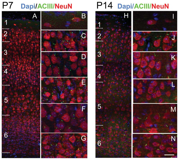
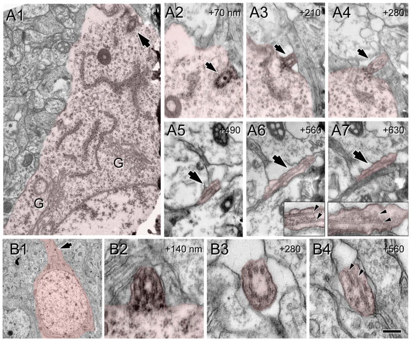
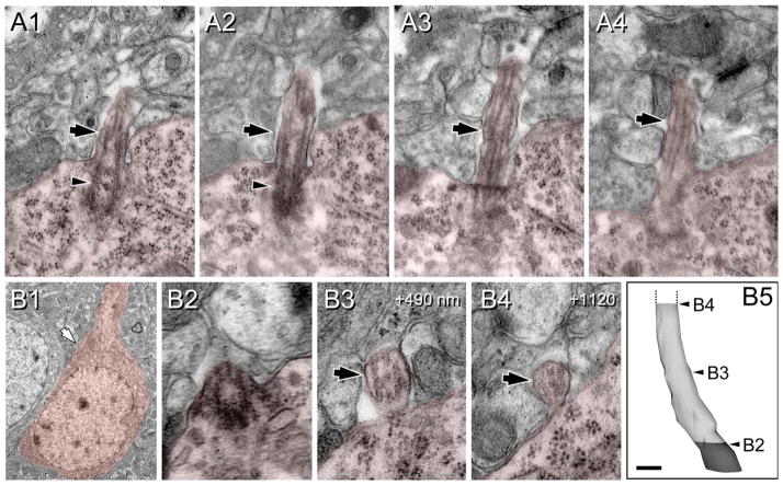
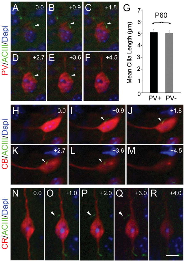
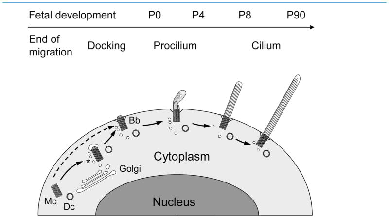
Neuronal primary cilia are not generally recognized, but they are considered to extend from most, if not all, neurons in the neocortex. However, when and how cilia develop in neurons are not known. This study used immunohistochemistry for adenylyl cyclase III (ACIII), a marker of primary cilia, and electron microscopic analysis to describe the development and maturation of cilia in mouse neocortical neurons. Our results indicate that ciliogenesis is initiated in late fetal stages after neuroblast migration, when the mother centriole docks with the plasma membrane, becomes a basal body, and grows a cilia bud that we call a procilium. This procilium consists of a membranous protrusion extending from the basal body but lacking axonemal structure and remains undifferentiated until development of the axoneme and cilia elongation starts at about postnatal day 4. Neuronal cilia elongation and final cilia length depend on layer position, and the process extends for a long time, lasting 8-12 weeks. We show that, in addition to pyramidal neurons, inhibitory interneurons also grow cilia of comparable length, suggesting that cilia are indeed present in all neocortical neuron subtypes. Furthermore, the study of mice with defective ciliogenesis suggested that failed elongation of cilia is not essential for proper neuronal migration and laminar organization or establishment of neuronal polarity. Thus, the function of this organelle in neocortical neurons remains elusive.


Figures

Figure 1
Figure 1
Expression of adenylyl cyclase III during fetal and postnatal cortical development. A: Maximum intensity projection of a z-stack of layer 3 pyramidal neurons in the neocortex of a P90 mouse. Cilia were immunostained with ACIII (green), basal bodies with pericentrin (red), neuronal somata with NeuN (purple), and nuclei with DAPI (blue). B: Western blot detection of ACIII from mouse cortical lysates from embryonic day 11.5 (E11.5) to young adulthood (~P60). The upper blot for ACIII revealed a band close to the predicted MW of unglycosylated ACIII (MW of ~125 kDa). Generally, ACIII expression significantly increases between P0 and P21. At P60, there is a decrease in the intensity of the ACIII signal. β-Actin (lower blot) was used as a loading control. C: Protein lysates of P90 olfactory epithelium (OE) or frontal cortex (FC) were separated by Western blot and probed for ACIII. Very strong expression of ACIII is detected at ~190–200 kDa, which has been shown to reflect high levels of glycosylated ACIII (bracket with asterisk). This higher MW signal was absent in FC sample that revealed only a lower MW signal for ACIII (lower bracket). Scale bar = 10 μm.
Figure 2
Figure 2
Basal bodies are more prevalent in the deeper cortical plate (CP) at E16.5. E13.5 embryos electroporated with cDNA encoding GFP, killed and fixed for immuno-EM at E16.5. A: DAB immunostaining with anti-GFP antibodies shows GFP+ cells (arrows) with leading and trailing processes in both the deeper layer and the upper layer of the CP. B: Higher magnification of the upper CP showing a cell (arrow) with a typical migrating profile. C: Bar graph shows a summary of the position of centrioles/basal bodies that were identified in deeper and upper layers of the CP docked (to the plasma membrane) or undocked. In deep CP, ~70% were docked (mostly in cells that were GFP). In upper CP, most centrioles (~90%) were undocked. D–I: Electron micrographic examples of centrioles and basal bodies in deep (D–G) or upper (H,I) CP. D1: A docked basal body in a GFP cell (boxed area is shown in D2). GFP immunoprecipitate is visible in the upper left cell (arrow). E: A docked basal body with a small axoneme extension (arrowhead). F1: Another example of a docked basal body in a GFP cell. The boxed region is shown in F2 and an adjacent section (AS) in F3. G1,3: Adjacent sections of a GFP+ cell (arrows point to GFP precipitate) with a leading process. Boxed regions in G1,3 are magnified in G2,4, respectively. H1: A GFP+ cell in the upper CP with undocked centrioles (boxed area is magnified in H2, which shows two centrioles (arrowheads). I1: Another example of a GFP+ cell in the upper CP. The boxed area in I1 is enlarged in I2, with an adjacent section shown in I3. Scale bars = 50 μm in A; 2 μm in D1,E,F1,G1,H1,I1; 0.5 μm in D2,F2,G2 H2.
Figure 3
Figure 3
Procilia in the fetal cortical plate at E16.5. Cells of interest and procilia are shaded to help identification. A: Serial sections (70 nm thick) illustrating an undocked mother centriole with an attached vesicle located adjacent to the cell membrane (asterisks). Inset in A1 shows the daughter centriole found in adjacent sections and (G) indicates Golgi cisterns around the centrosome. A2: A membranous centriolar protrusion, consistent with a procilium budding into the vesicle. This vesicular attachment is predictably the step before docking of the mother centriole to the cell membrane. B: Docked basal body with a protruding small procilium. B1: Low magnification image of the cell studied. Boxed area is magnified in B2. B2: Detail of the mother centriole (arrow) and the daughter centriole (arrowhead) located between the nucleus and the pial oriented process. B3–6: Serial sections (70 nm thick) illustrating the short procilium (~0.1 μm long) protruding from the attached mother centriole (arrow). C: Docked basal body with developed procilium. C1: Low magnification image of the cell analyzed showing the procilium (~0.3 μm long; arrow) protruding outside the cell. Inset shows an adjacent section illustrating the centriole surrounded by Golgi cisterns. C2–5: Serial sections (70 nm thick) illustrating the extent and mushroom shape of the procilium (arrow). Scale bar = 0.25 μm in C5 (applies to A,B3–6,C2–5); 3.6 μm for B1; 0.5 μm for B2; 0.54 μm for C1. [Color figure can be viewed in the online issue, which is available at wileyonlinelibrary.com.]
Figure 4
Figure 4
Elongation of neuronal cilia over several postnatal weeks. A–E: Examples of basal bodies (pericentrin+; red; example indicated by red arrowhead) and cilia (ACIII+; green; example indicated by green arrowhead) for the indicated ages. F: Bar graph shows the average ± SEM of cilia length (μm) in the neocortical layers at the indicated ages. Because it was difficult to differentiate neocortical lamina at early ages, average lengths for layers 2–4 and layers 5–6 were pooled for P0, whereas at P4 layers 3 and 4 were also pooled. G: Graphic representation of cilia elongation across age in the cortical layers. Mean values of length are used (SEM values are omitted for clarity but can be found in Table 2). H: Comparison of cilia length in upper layers (2–4; UL) and deep layers (5–6; DL) of the neocortex in male and female P14 mice did not show significant effect of gender. Scale bar = 2.5 μm.
Figure 5
Figure 5
Distribution of procilia in the neocortex at P0 and P4. A: Low-magnification view of the P0 neocortex immunostained for ACIII (green) and NeuN (red) and counterstained with DAPI (blue). NeuN is expressed in some cells of layers 2–5, scarce cells in layer 6, and intensely by some cells in the subplate (Sp). B–D: Details of the neocortex illustrating approximate layers 1–4 (L1, L2–4) in B, layers 5 and 6 (L5–6) in C, and the Sp in D. ACIII+ specks were found in all layers, with rare longer, rod-shaped cilia. E: Low-magnification view of the P4 neocortex immunostained for ACIII and NeuN and counterstained with DAPI. F–K: High-magnification details of the cortical layers from layer 1 to the Sp. NeuN is still not fully expressed by all neurons, and layer 2 (G) and deep layer 6 (J) have few stained neurons. In contrast, the subplate (K) shows large neurons intensely stained with NeuN. Overall, intense ACIII+ puncta are predominant, although scattered longer cilia can be found in all layers (arrows) and particularly in some neurons in layer 5 (I) and in the Sp (K). Scale bar = 10 μm in K (applies to F–K); 15 μm for A; 8 μm for B–D; 30 μm for E.
Figure 6
Figure 6
Heterogeneity of procilia at P0 in the upper cortical plate. Cells and procilia of interest are shaded to help with identification. A1: Low-magnification view of a cell with neuronal morphology in layer 2; arrow indicates the position of the docked centriole (basal body). A2 shows higher magnification of the basal body (Bb) and part of the adjacent daughter centriole (C). Note the lack of procilium (arrow) that was also absent in adjacent serial sections. B: A procilium in a cell with neuronal morphology in layer 2. B1: Low-magnification view of the cell with an arrow indicating the location of the procilium. B2: Cross-section of the basal body showing microtubular doublets (arrows) in the transitioning basal body/axonemal rudiment. B3–6: Serial sections (70 nm) through the procilium show the lack of axoneme but the presence of a tubular/vesicular network (arrows) inside the procilium. B7: 3D reconstruction of the procilium (white) and basal body (gray). C: Procilium in a cell with neuronal morphology. C1: Low-magnification view of a cell with an arrow indicating the location of the procilium. C2: Transition between the basal body (arrowhead) and procilium. C3–5: Serial sections illustrate a short procilium lacking microtubules and containing some vesicular structures (arrows). C6: 3D reconstruction of the basal body (gray) and the procilium (white) indicating the levels of C2–5 sections. D: Serial sections showing a procilium growing inside the cytoplasm in a cell in the upper cortical plate. D1: The basal body (arrowhead) and an adjacent vesicle (small arrow). D2: The basal body (arrowhead) and the budding procilium with a vesicle attached (large arrows). D3,4: The procilium is surrounded by the cell membrane and reaching the extracellular space in contact with an adjacent cell (discontinuous white line). Vesicles in and around the procilium are indicated with arrows. D5: Final section containing the procilium (asterisk). D6: Schematic of the procilia (Pc) growing inside the cytoplasm. Bb, basal body; V, vesicles; P, polyribosomes. Scale bar = 90 nm in D6 (applies to D1–5); 4 μm for A1; 0.18 μm for A2; 3 μm for B1; 70 nm for B2–6,C2–5; 0.15 μm for B7; 2 μm for C1; 0.1 μm for C6.
Figure 7
Figure 7
Procilia in the deep cortical plate at P0. Cells and procilia of interest are shaded to help with identification. A1: Low-magnification view of a cell with neuronal morphology bearing a procilium (arrow) in its pial aspect. A2–6: Serial sections of the procilium from the cell in A1. A2: The transition between the basal body (arrowhead) to the procilium. The procilium lacks an axoneme, but some microtubule-like structures are detectable (arrows in A2–6). A7: 3D reconstruction of the basal body (gray) and the (incomplete) procilium (white) indicating the relative positions of sections A2 and A6. B: Procilium in the deep cortical plate with “cabbage-like” morphology. B1: Low-magnification image of the cell with neuronal morphology bearing the procilium. B2,3: Serial micrographs showing the basal body (arrowhead) and the procilium with membrane foldings and multiple vesicles (arrows). C: Another example of a procilium containing multiple vesicles (arrows) and occasional tubular structures (small arrow). Vesicles (V) were also frequent around the basal body (arrowhead in C1). D: A cilium with axoneme in a cell with neuronal morphology in the subplate. D1: Low-magnification image of the cell bearing the cilium (arrow). D2–5: Selected serial oblique sections of the cilium illustrating the presence of parallel microtubules (arrows) forming the axoneme. The distance (nanometers) between sections is indicated at upper right. Surprisingly, the microtubules are more visible in distal sections (D4,5) than in proximal ones (D2,3). D6: 3D reconstruction of the cilium (basal body is not illustrated) showing the levels of sections D3–5. Scale bar = 0.16 μm in D5 (applies to D4,5); 2 μm for A1,B1; 90 nm for A2–6, 0.26 μm for A7; 0.18 μm for B2–3; 0.24 μm for C1,2; 1.8 μm for D1; 0.2 μm for D2,3; 0.25 μm for D6. [Color figure can be viewed in the online issue, which is available at wileyonlinelibrary.com.]
Figure 8
Figure 8
Procilia in P4 neocortex. Cells of interest and procilia are shaded to help with identification. Illustrated are examples of procilia lacking axonemes (A–C) and procilia located in the deep cortical plate with some microtubular structures but lacking a well-formed axoneme (D–F). A–C: Enlarged, apparently immature procilia, present both in the deep (A) and in the superficial (B,C) cortical plate. A1: Low-magnification view of a cell with neuronal morphology in the deep cortical plate; the arrow indicates the location of the procilium. A2,3: Serial longitudinal sections along the basal body (arrowhead) and procilium containing some microtubules (arrow). A multivesicular body (MB) and vesicles (V) could be found in the vicinity. B: Serial longitudinal sections along the basal body (arrowhead in B1) and stubby procilium containing a dark structure (arrowhead in B2). Vesicles (arrows in B2) were common close to the basal body. C1: Low-magnification view of a cell with neuronal morphology located in the deep cortical plate; the arrow indicates the location of the procilium. C2–6: Serial oblique sections through the basal body (arrowhead) and procilium (C3–6) containing vesicle-like structures (arrow in C5). C7: 3D reconstruction of the basal body (gray) and the cilium (white). D–F: Examples of procilia containing microtubule-like structures. D1: Low-magnification view of a cell with neuronal morphology located in the deep cortical plate; the arrow indicates the location of the procilium. D2–5: Serial sections (70 nm thick) through the procilium. D2: The basal body (arrowhead) and the adjacent Golgi apparatus (G). D3–5: Oblique sections through the procilium showing lack of axoneme with presence of scattered tubular structures (arrows). D6: 3D reconstruction of the basal body (gray) and the cilium (white). E1–6: Serial longitudinal sections along the basal body (arrowhead in E1) and the procilium containing disorganized microtubules (arrows). Golgi apparatus (G) vesicles were located close to the basal body. E6: 3D reconstruction of the basal body (gray) and the cilium (white). F1: Low-magnification view of the cell with neuronal morphology; the arrow indicates the location of the procilium. F2–5: Serial longitudinal sections along the basal body (arrowhead in F2) and the procilium that contains a few microtubules in a parallel arrangement (arrows), compatible with a developing axoneme. F5: 3D reconstruction of the basal body (gray) and the cilium (white). Scale bar = 0.2 μm in F5 (applies to F2–5); 2 μm for A1,C1,F1; 0.18 for A2,3,B1,2,C2–6,D6,E1–5; 0.25 for C7; 3 μm for D1; 0.16 μm for D2–5; 0.22 for E6. [Color figure can be viewed in the online issue, which is available at wileyonlinelibrary.com.]
Figure 9
Figure 9
ACIII+ cilia extend from neurons in all lamina of neocortex at P7 and P14. A: Low-magnification view of P7 neocortex immunostained for ACIII (green) and NeuN (red) and counterstained with DAPI (blue). B–G: High-magnification views of layers 1 (B), 2 (C), 3 (D), 4 (E), 5 (F), and 6 (G). Compared with P4 (Fig. 5), layers are more developed, neuropil is expanding, and cilia are longer and growing at similar rates in all layers (see Table 2). H–N: Low-magnification view of P14 neocortex immunostained for ACIII and NeuN and counterstained with DAPI. High-magnification views of layers 1 (I), 2 (J), 3 (K), 4 (L), 5 (M), and 6 (N) are shown at right. Note the expansion of the neuropil and more elongated appearance of cilia in all layers. At this age, cilia lengths reach ~70–90% of maximal lengths (see Table 2). Scale bar = 20 μm in N (applies to I–N); 70 μm for A; 24 μm for B–G; 70 μm for H.
Figure 10
Figure 10
Ultrastructure of cilia at P8. Cells of interest and procilia are shaded to help with identification. A: Serial micrographs of a cilium at P8. A1: Panoramic view of the cytoplasm and initial apical dendrite of a pyramidal neuron with the basal body attached to the membrane (arrow). G, Golgi apparatus. A2–7: Serial sections of the cilium (arrow) protruding outside of the cell. Well-formed and structured microtubules in the proximal segment (insets in A6,7) appear disorganized distally (inset in A7). Numbers in upper right corner indicate the Z distance between sections in nanometers. B: Serial sections of a cilium from a pyramidal neuron at P8. B1: Panoramic view of the pyramidal cell. Cilium location is indicated (arrow). B2–4: Serial micrographs of the cilia from neuron in B1. B2 illustrates the transition between the basal body and the cilium, and B3,4 are transverse and oblique sections through the cilium showing a straight morphology and a well-developed axoneme along the available length of 0.56 μm. Vesicles were less frequent at this age. Scale bar = 0.12 μm in B4 (applies to B2–4); 0.5 μm for A1; 0.4 μm for A2–7; 5 μm for B1. [Color figure can be viewed in the online issue, which is available at wileyonlinelibrary.com.]
Figure 11
Figure 11
Ultrastructure of cilia at P60. Cells of interest and procilia are shaded to help with identification. A: Serial sections along the basal body (arrowhead) and proximal segment (arrow) of a neuronal cilium. Notice the well-developed microtubules. B: Serial transverse sections of a pyramidal cell cilium (incomplete). B1: Panoramic view of a pyramidal neuron. Location of the cilium is indicated (arrow). B2–4: Details of transverse sections through the cilium of the pyramidal neuron in B1. B2: Transition basal body-cilium. B3: Middistance of the series. B4: Final section at about 1.2 μm from the origin. Note the rounded shape of the ciliar membrane and the well-developed axoneme along the available length. B5: 3D reconstruction of the basal body (gray) and the (incomplete) cilium (white). Positions of images B2–4 are indicated. Numbers in upper right corner indicate the Z distance to the cilium origin in nanometers. Scale bar = 0.18 μm in B5; 0.17 μm for A; 1.5 μm for B1; 0.1 for B2–4. [Color figure can be viewed in the online issue, which is available at wileyonlinelibrary.com.]
Figure 12
Figure 12
Different interneuron subtypes in neocortex extend cilia. Examples of ciliated interneurons in the neocortex of P60 mice. A–F: An ACIII+ (arrow) cilium extending out of a parvalbumin (PV)-positive cell. G: Bar graph shows cilia length between PV+ and neighboring PV cells were comparable (~5 μm). H–M: Example of a calbindin (CB)-positive interneuron with an ACIII+ cilium (arrow). N–R: A calretinin (CR)-positive interneuron extending an ACIII+ cilium (arrow). Interestingly, some bipolar CR+ cells extended their cilia from the proximal part of the ascending (pial-oriented) dendrite, as illustrated. Numbers in the right upper corner of images indicate z-step increments in micrometers. Scale bar = 7.5 μm in R (applies to N–R); 12 μm for A–F; 10 μm for H–M.
Figure 13
Figure 13
Mutants lacking cilia show normal gross cytoarchitecture. A: Panoramic view of the neocortex of ΔStumpy mutant mice immunostained for NeuN (green) and counterstained with DAPI (blue). The boxed areas in A represent higher magnification views of layers 2–3 (B) and 5 (C). In spite of the cortical compression resulting from hydrocephalus, ΔStumpy mice lacking cilia exhibit normal polarization in pyramidal neurons, with well-formed and oriented apical dendrites (arrows in B,C). D,E: Panoramic view of the cortical plate (cp) of P10 control (D1,2) and ΔStumpy mice (E1,2) immunostained for Cux1 (upper layer neurons marker; green) and Foxp2 (deeper layer neurons marker; red). Stratification of cortical layers was also grossly preserved in mice lacking cilia. D2 and E2 have had the DAPI channel removed.
Figure 14
Figure 14
Model of ciliogenesis stages in mouse neocortical neurons. We propose the following model. Migrating neurons do not bear cilia; rather, their mother centriole (Mc) and daughter centriole (Dc) are free in the cytoplasm. Once cells terminate migration and reach their appropriate lamina, the mother centriole attaches a vesicle, likely from the Golgi apparatus, buds a very short procilium and docks to the plasma membrane. It is also possible that the mother centriole docks directly to the plasma membrane without vesicle attachment as indicated by the dashed arrow. Docking to the membrane involves developing specific structures such as transition filaments, and the mother centriole will become a basal body (Bb), frequently surrounded by vesicles (asterisk). This basal body will grow the procilium, a membranous expansion about 0.5–2 μm in length, whose main feature is the lack of proper axoneme and that typically contains vesicles, short and disorganized tubular structures, and electron-dense diffuse content. This procilium does not display typical axonemal characteristics until ~P8, although axonemal growth seems to start at ~P0 in some subplate cells and could start ~P4 in some populations of neurons that showed early elongation of cilia (e.g., some layer 5 neurons). Overall, cilia will take weeks to elongate fully toward a peak at ~P60–P90, with some differences between layers.

References

    1. Airaksinen MS, Eilers J, Garaschuk O, Thoenen H, Konnerth A, Meyer M. Ataxia and altered dendritic calcium signaling in mice carrying a targeted null mutation of the calbindin D28k gene. Proc Natl Acad Sci U S A. 1997;94:1488–1493. - PMC - PubMed
    1. Amador-Arjona A, Elliott J, Miller A, Ginbey A, Pazour GJ, Enikolopov G, Roberts AJ, Terskikh AV. Primary cilia regulate proliferation of amplifying progenitors in adult hippocampus: implications for learning and memory. J Neurosci. 2011;31:9933–9944. - PMC - PubMed
    1. Anastas SB, Mueller D, Semple-Rowland SL, Breunig JJ, Sarkisian MR. Failed cytokinesis of neural progenitors in citron kinase-deficient rats leads to multiciliated neurons. Cereb Cortex. 2011;21:338–344. (Epub 2010, Jun 4) - PubMed
    1. Barzi M, Berenguer J, Menendez A, Alvarez-Rodriguez R, Pons S. Sonic-hedgehog-mediated proliferation requires the localization of PKA to the cilium base. J Cell Sci. 2010;123:62–69. - PubMed
    1. Bearzatto B, Servais L, Roussel C, Gall D, Baba-Aissa F, Schurmans S, de Kerchove d’Exaerde A, Cheron G, Schiffmann SN. Targeted calretinin expression in granule cells of calretinin-null mice restores normal cerebellar functions. FASEB J. 2006;20:380–382. - PubMed

Publication types

MeSH terms