. 2011 Dec 9;286(49):42435-42445.
doi: 10.1074/jbc.M110.212845. Epub 2011 Oct 21.

Phosphoinositide binding differentially regulates NHE1 Na+/H+ exchanger-dependent proximal tubule cell survival

Affiliations

Phosphoinositide binding differentially regulates NHE1 Na+/H+ exchanger-dependent proximal tubule cell survival

Bassam G Abu Jawdeh et al. J Biol Chem. .

Abstract

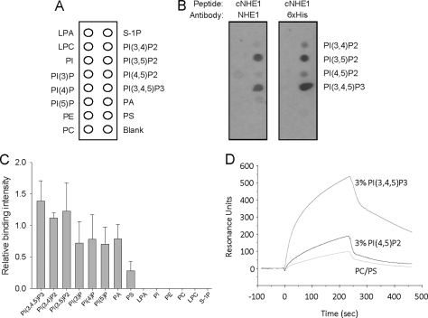
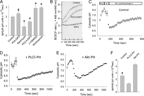
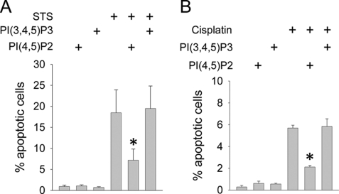
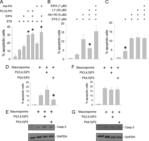
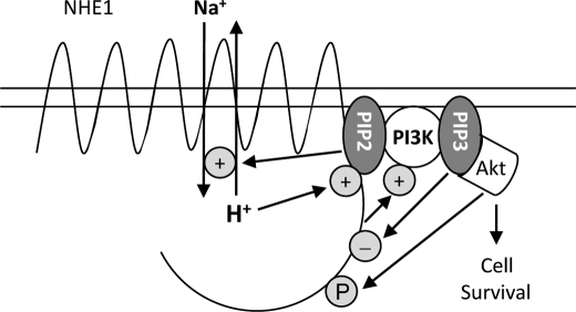
Tubular atrophy predicts chronic kidney disease progression, and is caused by proximal tubular epithelial cellcaused by proximal tubular epithelial cell (PTC) apoptosis. The normally quiescent Na(+)/H(+) exchanger-1 (NHE1) defends against PTC apoptosis, and is regulated by PI(4,5)P(2) binding. Because of the vast array of plasma membrane lipids, we hypothesized that NHE1-mediated cell survival is dynamically regulated by multiple anionic inner leaflet phospholipids. In membrane overlay and surface plasmon resonance assays, the NHE1 C terminus bound phospholipids with low affinity and according to valence (PIP(3) > PIP(2) > PIP = PA > PS). NHE1-phosphoinositide binding was enhanced by acidic pH, and abolished by NHE1 Arg/Lys to Ala mutations within two juxtamembrane domains, consistent with electrostatic interactions. PI(4,5)P(2)-incorporated vesicles were distributed to apical and lateral PTC domains, increased NHE1-regulated Na(+)/H(+) exchange, and blunted apoptosis, whereas NHE1 activity was decreased in cells enriched with PI(3,4,5)P(3), which localized to basolateral membranes. Divergent PI(4,5)P(2) and PI(3,4,5)P(3) effects on NHE1-dependent Na(+)/H(+) exchange and apoptosis were confirmed by selective phosphoinositide sequestration with pleckstrin homology domain-containing phospholipase Cδ and Akt peptides, PI 3-kinase, and Akt inhibition in wild-type and NHE1-null PTCs. The results reveal an on-off switch model, whereby NHE1 toggles between weak interactions with PI(4,5)P(2) and PI(3,4,5)P(3). In response to apoptotic stress, NHE1 is stimulated by PI(4,5)P(2), which leads to PI 3-kinase activation, and PI(4,5)P(2) phosphorylation. The resulting PI(3,4,5)P(3) dually stimulates sustained, downstream Akt survival signaling, and dampens NHE1 activity through competitive inhibition and depletion of PI(4,5)P(2).


Figures

FIGURE 1.
FIGURE 1.
NHE1 binds multiple membrane lipids. Purified and renatured His6-tagged cNHE1 was incubated with membrane phospholipids spotted on nitrocellulose membranes (A), and probed for binding with either anti-NHE1 or anti-His antibodies (B). C, quantitation of cNHE1 binding by densitometry, as determined by anti-NHE1 peptide antibody detection. Data are normalized to PI(4,5)P2 binding (defined as 1.0 in each experiment). D, phospholipid binding to the immobilized cNHE1 peptide by surface plasmon resonance. 70% PC, 30% PS (50 μm) served as a control for inner leaflet phospholipids. Experimental groups include 50 μm C16-PI(4,5)P2 or 50 μm C16-PI(3,4,5)P3 (3% (w/w) in 70% PC, 30% PS vesicles). Phospholipids were dispersed by sonication and passage through an extruder. Analyte suspensions were flowed at 5 μl/min for 4 min to establish association, followed by a 4-min perfusion with buffer only, for dissociation.
FIGURE 2.
FIGURE 2.
NHE1 binds phosphoinositides through electrostatic interactions. Association of diC8-PI(4,5)P2 and diC8-PI(3,4,5)P3 with wild-type and mutant cNHE1 peptides (mutated residues are shown in A), as measured by surface plasmon resonance (B). Mutant cNHE1 binding to phospholipids, by membrane overlay assays (C). In control experiments binding to phosphatidylinositol monophosphates in the absence of cNHE1 peptides was occasionally observed (not shown), indicating a lack of binding specificity to these sites.
FIGURE 3.
FIGURE 3.
Effect of pH on NHE1-phosphoinositide binding. NHE1 association with diC8-PI(4,5)P2 (A) and diC8-PI(3,4,5)P3 (B) at varying pH values within the physiologic range. Dissociation rates were not significantly different between pH conditions (not shown). Note that the initial, steep curves are a result of bulk solute shift, rather than specific binding. C, representative epifluorescence images of RFP-NHE1-transfected and untransfected cells and corresponding TIRF images of BODIPY fluorescence. Cytosolic pH was clamped at pH 6.5, 7.0, and 7.5 following exposure to nigericin (10 μm, 15 min) in 100 mm KH2PO4 + 30 mm NaCl buffer. D, energy transfer efficiency between PI(4,5)P2-BODIPY (donor) and RFP-NHE1 (acceptor) measured by TIRF microscopy. Data are expressed as mean ± S.E. from three experiments, 8–10 pairs of cells within the same microscopic field per experiment.
FIGURE 4.
FIGURE 4.
PI(4,5)P2, PI(3,4,5)P3, and NHE1 co-localize to lateral membrane domains. LLC-PK1 cells were polarized on permeable supports and transiently transfected with GFP-tagged PLCδ (A–C and G–J) or Akt (D–F and K–N) PH domain peptide cDNAs. NHE1 distribution was determined immunocytochemically with anti-NHE1, followed by Texas Red-conjugated anti-rabbit IgG (B, E, H, and L). Apical-basolateral junctions were labeled with ZO-1 antibodies, followed by AF488-conjugated goat anti-rat IgG (I and M, demarcated with arrowheads for clarity). Phosphoinositide, NHE1, and ZO-1 localization were detected in paraformaldehyde-fixed cells by confocal microscopy, and displayed in the X-Y (A–F) and X-Z plane (G–N).
FIGURE 5.
FIGURE 5.
PI(4,5)P2 and PI(3,4,5)P3 differentially regulate NHE1-dependent Na+/H+ exchange. A, LLC-PK1 cells were preincubated with metabolically stable (phosphatase-resistant) diC8-PI(4,5)P2 or diC8-PI(3,4,5)P3, PI(4,5)P2 inhibitor neomycin, or PI 3-kinase inhibitors LY-294002 (20 μm, 30 min, 37 °C) or wortmannin (100 nm, 30 min, 37 °C). Maximum EIPA (1 μm)-inhibitable Na+/H+ exchange was determined in response to cytosolic acidification following NH4Cl washout. Cytosolic pH was determined by BCECF fluorescence, and measured by spectrofluorimetry. Results from four to six experiments per condition are shown. *, p < 0.05 and §, p = 0.12 compared with control group by analysis of variance on ranks with the Dunnett post hoc test. B, representative pH recovery tracing from one experiment in PI(3,4,5)P3-loaded versus control cells. C–E, NHE1-dependent (EIPA (1 μm)-inhibitable) Na+/H+ exchange was measured in individual, BCECF-loaded LLC-PK1 cells by fluorescence microscopy following NH4Cl washout (W in C). Mean values from three experiments, 35–50 cells per experiment, are shown for GFP-transfected (C), GFP-tagged PLCδ-PH-expressing (D), and GFP-tagged Akt-PH-expressing (E) cells. Maximum rates of EIPA-inhibitable Na+/H+ exchange are summarized in F. *, p < 0.05 compared with other groups by analysis of variance.
FIGURE 6.
FIGURE 6.
PI(4,5)P2 and PI(3,4,5)P3 differentially regulate apoptosis. LLC-PK1 cells were maintained on permeable supports, and pre-treated by incorporation of metabolically stable diC8-PI(4,5)P2 or diC8-PI(3,4,5)P3. Cells were then induced to undergo apoptosis with staurosporine (STS, 1 μm, 5 h, in panel A) or cisplatin (25 μm, 18 h, 37 °C, in panel B). Apoptosis was quantified by TUNEL. Note the difference in y-axis scale between histograms. *, p < 0.05 compared with staurosporine-only or cisplatin-only groups by analysis of variance.
FIGURE 7.
FIGURE 7.
PI(4,5)P2 and PI(3,4,5)P3 differentially regulate NHE1-dependent cell survival. LLC-PK1 cells (A), as well as wild-type (B, D, and E) and NHE1-null mouse proximal tubule cell lines (C, F, and G) were cultured on permeable supports. LLC-PK1 cells underwent PI(4,5)P2 or PI(3,4,5)P3 sequestration by overexpressing GFP-tagged PLCδ-PH or Akt-PH peptide constructs, respectively (A), or preincubation with PI 3-kinase inhibitor LY-294002, Akt inhibitor Akt VIII, or NHE1 inhibitor EIPA (B and C). Mouse PTC were loaded with PI(4,5)P2 or PI(3,4,5)P3 vesicles (D–G). All groups were induced to undergo apoptosis with staurosporine (STS, 1 μm, 5 h, 37 °C). Apoptosis was determined by TUNEL assay (A–D and F) or detection with antibodies that recognize the active, 17 kDa, cleaved caspase-3 by immunoblot analysis (E and G, upper panels). Blots were stripped and re-probed for GAPDH as a loading control (E and G, lower panels). *, p < 0.05 compared with STS alone by analysis of variance on ranks and the Dunnett post hoc analysis in A; *, p < 0.05 compared with all other groups by analysis of variance in B–D.
FIGURE 8.
FIGURE 8.
Schematic diagram summarizing the role of phosphoinositides on regulation of NHE1-dependent cell survival. In response to apoptotic stress, NHE1 is initially stimulated by PI(4,5)P2 binding to mediate cytoprotection. NHE1 then indirectly activates PI 3-kinase to phosphorylate PI(4,5)P2. The resulting PI(3,4,5)P3 dually stimulates sustained downstream Akt survival signaling, and negatively feeds back to dampen NHE1 activity through competitive inhibition, as well as depletion of PI(4,5)P2 pools.

References

    1. Coresh J., Selvin E., Stevens L. A., Manzi J., Kusek J. W., Eggers P., Van Lente F., Levey A. S. (2007) JAMA 298, 2038–2047 - PubMed
    1. Collins A. J., Foley R. N., Herzog C., Chavers B. M., Gilbertson D., Ishani A., Kasiske B. L., Liu J., Mau L. W., McBean M., Murray A., St P. W., Guo H., Li Q., Li S., Li S., Peng Y., Qiu Y., Roberts T., Skeans M., Snyder J., Solid C., Wang C., Weinhandl E., Zaun D., Arko C., Chen S. C., Dalleska F., Daniels F., Dunning S., Ebben J., Frazier E., Hanzlik C., Johnson R., Sheets D., Wang X., Forrest B., Constantini E., Everson S., Eggers P. W., Agodoa L. (2010) Am. J. Kidney Dis. 55, S1-S7 - PMC - PubMed
    1. Risdon R. A., Sloper J. C., De Wardener H. E. (1968) Lancet 2, 363–366 - PubMed
    1. Schainuck L. I., Striker G. E., Cutler R. E., Benditt E. P. (1970) Hum. Pathol. 1, 631–641 - PubMed
    1. Bohle A., Muller G. A., Wehrmann M., Mackensen-Haen S., Xiao J. C. (1996) Kidney Int. 54, S2–S9 - PubMed

Publication types