. 2011 Dec 23;286(51):43717-43725.
doi: 10.1074/jbc.M111.292755. Epub 2011 Oct 21.

Junctophilin 1 and 2 proteins interact with the L-type Ca2+ channel dihydropyridine receptors (DHPRs) in skeletal muscle

Affiliations

Junctophilin 1 and 2 proteins interact with the L-type Ca2+ channel dihydropyridine receptors (DHPRs) in skeletal muscle

Lucia Golini et al. J Biol Chem. .

Abstract

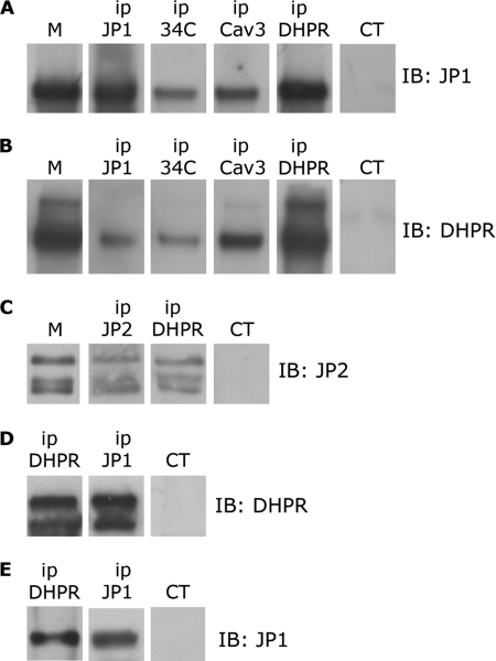
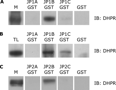
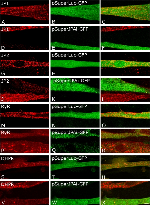
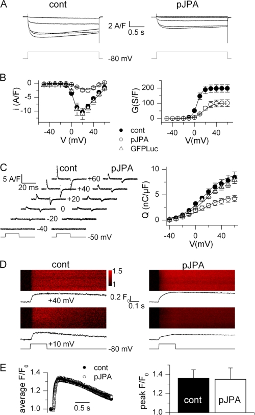
Junctophilins (JPs) anchor the endo/sarcoplasmic reticulum to the plasma membrane, thus contributing to the assembly of junctional membrane complexes in striated muscles and neurons. Recent studies have shown that JPs may be also involved in regulating Ca2+ homeostasis. Here, we report that in skeletal muscle, JP1 and JP2 are part of a complex that, in addition to ryanodine receptor 1 (RyR1), includes caveolin 3 and the dihydropyridine receptor (DHPR). The interaction between JPs and DHPR was mediated by a region encompassing amino acids 230-369 and amino acids 216-399 in JP1 and JP2, respectively. Immunofluorescence studies revealed that the pattern of DHPR and RyR signals in C2C12 cells knocked down for JP1 and JP2 was rather diffused and characterized by smaller puncta in contrast to that observed in control cells. Functional experiments revealed that down-regulation of JPs in differentiated C2C12 cells resulted in a reduction of intramembrane charge movement and the L-type Ca2+ current accompanied by a reduced number of DHPRs at the plasma membrane, whereas there was no substantial alteration in Ca2+ release from the sterol regulatory element-binding protein. Altogether, these results suggest that JP1 and JP2 can facilitate the assembly of DHPR with other proteins of the excitation-contraction coupling machinery.


Figures

FIGURE 1.
FIGURE 1.
Immunoprecipitation of DHPR, JP1, and JP2 from solubilized rabbit skeletal muscle and HEK293-T microsomes. A, 100 μg of proteins solubilized from rabbit skeletal muscle microsomes (M) were immunoprecipitated (ip) with antibodies against JP1, RyR (34C), caveolin-3 (Cav3), α1s-DHPR (DHPR), and a non-correlated control antibody (CT). After gel electrophoresis, proteins were transferred to a nylon membrane and incubated with anti-JP1 antibody. B, 100 μg of proteins solubilized from skeletal muscle microsomes were immunoprecipitated with antibodies against JP1, RyR, caveolin-3, and α1s-DHPR and a non-correlated control antibody. After gel electrophoresis, proteins were transferred to a nylon membrane and incubated with an antibody against α1s-DHPR. C, 100 μg of proteins solubilized from rabbit skeletal muscle microsomes were immunoprecipitated with antibodies against JP2, α1s-DHPR, and a non-correlated control antibody. After gel electrophoresis, proteins were transferred to a nylon membrane and incubated with anti-JP2 antibody. D, solubilized proteins from HEK293-T cells transfected with pEGFP-α1s-DHPR, pEGFP-β1a-DHPR, and pCDNA-JP1 were immunoprecipitated with antibodies against α1s-DHPR, JP1, and a non-correlated control antibody. Proteins were separated by gel electrophoresis, transferred to a nylon membrane, and incubated with an antibody against DHPR. E, solubilized proteins from HEK293-T cells transfected with pEGFP-α1s-DHPR, pEGFP-β1a-DHPR, and pCDNA-JP1 were immunoprecipitated with antibodies against α1s-DHPR, JP1, and a non-correlated control antibody. Proteins were separated by gel electrophoresis, transferred to a nylon membrane, and incubated with an anti-JP1 antibody.
FIGURE 2.
FIGURE 2.
Association of DHPR with JP1-B in skeletal muscle and in HEK293-T transfected cells. A, Western blot (IB) with an anti-α1s-DHPR antibody of solubilized rabbit skeletal muscle microsomes (M) and of proteins pulled down with GST-JP1-A, GST-JP1-B, GST-JP1-C, and GST alone, as negative control. DHPR, anti-α1s-DHPR antibody. B, Western blot with an anti-α1s-DHPR antibody of lysates of HEK293-T cells transfected with pEGFP-α1s-DHPR, pEGFP-β1a-DHPR, and pCDNA-JP1 (TL) and of proteins pulled down with GST-JP1-A, GST-JP1-B, GST-JP1-C, and GST alone, as negative control. C, Western blot with an anti-α1s-DHPR antibody of solubilized rabbit skeletal muscle microsomes and proteins pulled down with GST-JP2-A, GST-JP2-B, GST-JP2-C, and GST alone, as negative control.
FIGURE 3.
FIGURE 3.
Immunofluorescence analysis of expression inhibition of JP1 and JP2 in C2C12 cells. C2C12 cells were transfected with pSuperLuc-GFP as negative control (A–C, G–I, M–O, and S–U) or with pSuperJPAi-GFP to silence JP1 and JP2 expression (D-F, J–L, P–R, and V–X). C2C12 cells transfected with pSuperLuc-GFP were immunostained with antibodies against JP1 (A–C), JP2 (G–I), RyR (M–O), and α1s DHPR (S–U). C2C12 cells were transfected with pSuperJPAi-GFP were immunostained with antibodies against JP1 (D–F), JP2 (J–L), RyR (P–R), and α1s-DHPR (V–X).
FIGURE 4.
FIGURE 4.
DHPR-mediated Ca2+ entry, charge movement, and Ca2+ release in control and JP knockdown C2C12 myotubes. A, Ca2+ current traces obtained in a control myotube (cont, left) and in a pSuperJPAi-GFP-positive myotube (pJPA, right) in response to the depolarizing pulse protocol illustrated below. B, mean voltage dependence of the peak Ca2+ current density (left) and corresponding mean maximum conductance (right) in control (n = 5), pSuperJPAi-GFP-positive (n = 6), and pSuperLuc-GFP-positive (n = 5) myotubes. Superimposed lines were calculated from the average values of the parameters obtained from fitting the appropriate function to the individual series of data (see ”Experimental Procedures“). C, left, charge movement currents measured in a control and in a pSuperJPAi-GFP-positive myotube at the indicated values of membrane potential. Right, mean voltage dependence of charge density in control (n = 13), pSuperJPAi-GFP-positive (n = 9), and pSuperLuc-GFP-positive myotubes (n = 10). Superimposed lines were calculated from the average values of the parameters obtained from fitting a Boltzmann function to the individual series of data. D, line-scan images of the rhod-2 fluorescence taken from a control myotube (left) and from a pSuperJPAi-GFP-positive myotube (right). Vertical size corresponds to 100 μm. Myotubes were stimulated by a 200-ms-long voltage clamp depolarization from −80 mV to the indicated level. The average time course of change in rhod-2 fluorescence is shown underneath each line-scan image. E, left, average rhod-2 fluorescence transient elicited by a depolarizing pulse to +40 mV in nine control and seven pSuperJPAi-GFP-positive myotubes. Right, corresponding mean (± S.E.) peak amplitude of the rhod-2 transient in the two sets of myotubes.

References

    1. Franzini-Armstrong C. (2004) in Myology (Engel A. G., Franzini-Armstrong C. eds) pp. 232–256, McGraw-Hill, Inc., New York, NY
    1. Sorrentino V. (2011) Int. J Biochem. Cell Biol. 43, 1075–1078 - PubMed
    1. Rossi A. E., Dirksen R. T. (2006) Muscle Nerve 33, 715–731 - PubMed
    1. Takahashi M., Seagar M. J., Jones J. F., Reber B. F., Catterall W. A. (1987) Proc. Natl. Acad. Sci. U.S.A. 84, 5478–5482 - PMC - PubMed
    1. Leung A. T, Imagawa T., Campbell K. P. (1987) J. Biol. Chem. 262, 7943–7946 - PubMed

Publication types

LinkOut - more resources