. 2012 Apr;67(4):313-29.
doi: 10.1093/gerona/glr164. Epub 2011 Oct 21.

Liver-specific knockdown of IGF-1 decreases vascular oxidative stress resistance by impairing the Nrf2-dependent antioxidant response: a novel model of vascular aging

Affiliations

Liver-specific knockdown of IGF-1 decreases vascular oxidative stress resistance by impairing the Nrf2-dependent antioxidant response: a novel model of vascular aging

Lora C Bailey-Downs et al. J Gerontol A Biol Sci Med Sci. 2012 Apr.

Abstract

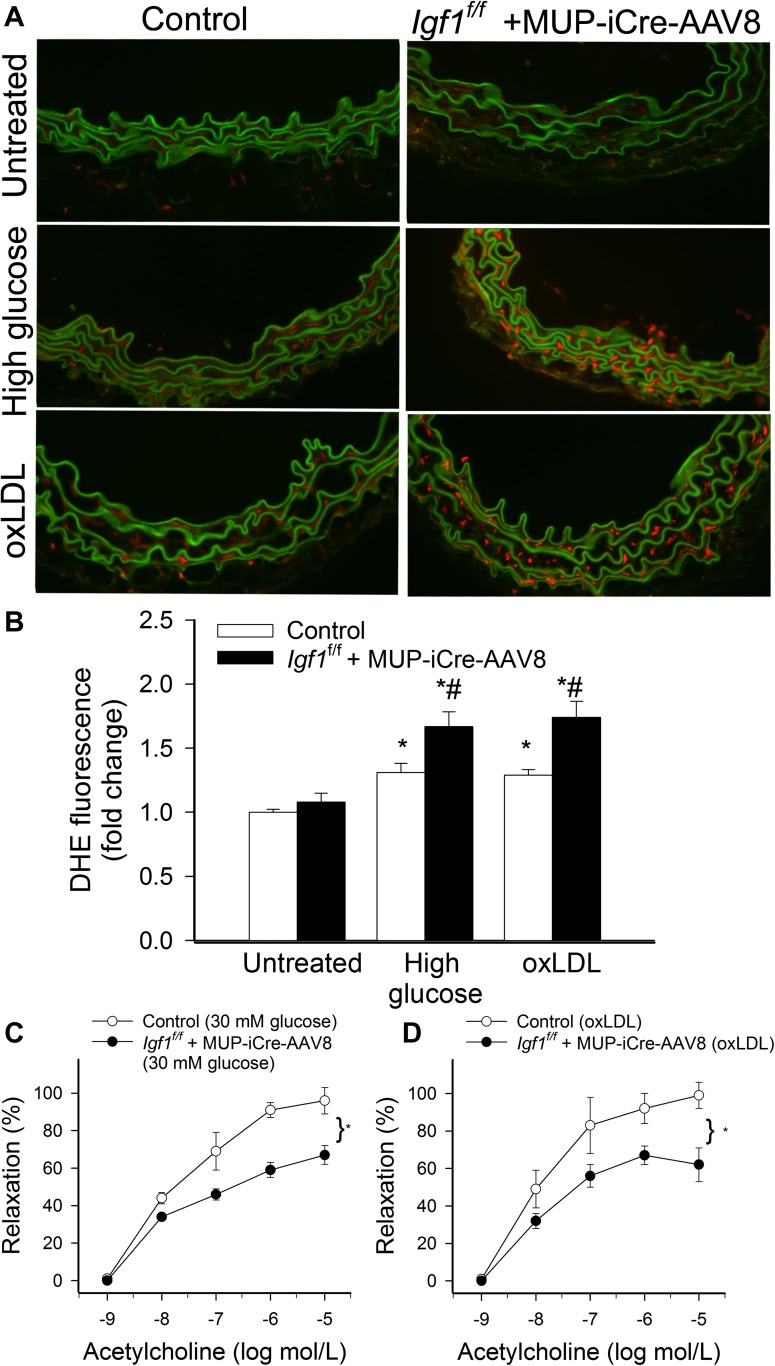
Recent studies demonstrate that age-related dysfunction of NF-E2-related factor-2 (Nrf2)-driven pathways impairs cellular redox homeostasis, exacerbating age-related cellular oxidative stress and increasing sensitivity of aged vessels to oxidative stress-induced cellular damage. Circulating levels of insulin-like growth factor (IGF)-1 decline during aging, which significantly increases the risk for cardiovascular diseases in humans. To test the hypothesis that adult-onset IGF-1 deficiency impairs Nrf2-driven pathways in the vasculature, we utilized a novel mouse model with a liver-specific adeno-associated viral knockdown of the Igf1 gene using Cre-lox technology (Igf1(f/f) + MUP-iCre-AAV8), which exhibits a significant decrease in circulating IGF-1 levels (~50%). In the aortas of IGF-1-deficient mice, there was a trend for decreased expression of Nrf2 and the Nrf2 target genes GCLC, NQO1 and HMOX1. In cultured aorta segments of IGF-1-deficient mice treated with oxidative stressors (high glucose, oxidized low-density lipoprotein, and H(2)O(2)), induction of Nrf2-driven genes was significantly attenuated as compared with control vessels, which was associated with an exacerbation of endothelial dysfunction, increased oxidative stress, and apoptosis, mimicking the aging phenotype. In conclusion, endocrine IGF-1 deficiency is associated with dysregulation of Nrf2-dependent antioxidant responses in the vasculature, which likely promotes an adverse vascular phenotype under pathophysiological conditions associated with oxidative stress (eg, diabetes mellitus, hypertension) and results in accelerated vascular impairments in aging.


Figures

Figure 1.
Figure 1.
A: Serum insulin-like growth factor (IGF)-1 protein levels in mice with hepatic IGF-1 knockdown (Igf1f/f + MUP-iCre-AAV8, initiated at 5 months of age and assessed at 11 months of age) and control (Igf-1f/f + MUP-eGFP-AAV8) mice. Data are mean ± SEM. *p < .05 versus control (n = 20 in each group). B: Acetylcholine induced relaxation of aorta rings isolated from IGF-1–deficient and control mice. Data are mean ± SEM (n = 4 in each group). C: Production of H2O2 in the aortas of IGF-1–deficient and control mice as assessed by Amplex Red/horseradish peroxidase assay. Data are mean ± SEM (n = 6 in each group). GSH (D) and ascorbate (E) content, determined using high-performance liquid chromatography electrochemical detection, in the aorta of IGF-1–deficient and control mice. Data are mean ± SEM (n = 10 in each group).
Figure 2.
Figure 2.
Expression of Nrf2 mRNA (A) and protein (B; upper panel: representative Western blots, lower panel: bar graphs are normalized densitometric data) in the aortas of insulin-like growth factor (IGF)-1–deficient (Igf1f/f + MUP-iCre-AAV8) and control (Igf-1f/f + MUP-eGFP-AAV8) mice, assessed by quantitative real-time reverse transcription–polymerase chain reaction and Western blotting, respectively. Data are mean ± SEM. *p < .05 versus control (n = 5–9 in each group).
Figure 3.
Figure 3.
Quantitative real-time reverse transcription–polymerase chain reaction data showing messenger RNA expression of Gclc (A), Nqo1 (B), Hmox1 (C), and catalase (D) in the aortas of insulin-like growth factor (IGF)-1 deficient (Igf1f/f + MUP-iCre-AAV8) and control (Igf-1f/f + MUP-eGFP-AAV8) mice. EF: Protein expression of GCLC (E) and NQO1 (F) in the aortas of IGF-1–deficient and control mice. Upper panels: representative Western blots, lower panel: bar graphs are normalized densitometric data. Data are mean ± SEM. *p < .05 versus control (n = 9 in each group).
Figure 4.
Figure 4.
A: Reporter gene assay showing the effects of recombinant insulin-like growth factor (IGF)-1 (200 ng/mL) on Nrf2/ARE reporter activity in cultured primary human coronary arterial endothelial cells (CAECs). Cells were transiently cotransfected with ARE-driven firefly luciferase and CMV-driven renilla luciferase constructs followed by IGF-1 treatment. Cells were then lysed and subjected to luciferase activity assay. After normalization relative luciferase activity was obtained from four to six independent transfections. Data are mean ± SEM. *p < .05. BD: Effect of IGF-1 on messenger RNA expression of Nqo1, Hmox1, and Gclc in cultured primary CAECs. Data are mean ± SEM (n = 5 in each group). The effects of IGF-1 were significant (p < .05) for each target. E: Effect of IGF-1 on Nrf2/ARE reporter activity in CAECs transfected with plasmids expressing the wild-type human Akt1 (pMEV2HA-AKT1-WT) or a dominant negative mutant form of Akt1 (pMEV2HA-AKT1-K179A, pMEV2HA-AKT1-AA). Data are mean ± SEM (n = 6–8 in each group). *p < .05 versus untreated control, #p < .05 versus respective wild type.
Figure 5.
Figure 5.
Quantitative real-time reverse transcription–polymerase chain reaction data showing H2O2 (1 μmol/L) and high glucose (30 mmol/L)–induced changes in messenger RNA expression of Gclc (A), Nqo1 (B), Hmox1 (C), and catalase (D) in cultured aorta segments isolated from insulin-like growth factor (IGF)-1–deficient (Igf1f/f + MUP-iCre-AAV8) and control (Igf-1f/f + MUP-eGFP-AAV8) mice. E: H2O2-induced increases in Nrf2-binding activity in nuclear extracts from aorta segments of Igf1f/f + MUP-iCre-AAV8 and control mice. Data are mean ± SEM (n = 9 in each group). *p < .05 versus untreated, #p < .05 versus Igf-1f/f + MUP-eGFP-AAV8.
Figure 6.
Figure 6.
A: Reporter gene assay showing the effects of oxLDL (40 μg/mL) on Nrf2/ARE reporter activity in primary human coronary arterial endothelial cells (CAECs) cultured in the presence of sera derived from Igf1f/f + MUP-iCre-AAV8 or control mice (n = 18–20 in each group). Data are mean ± SEM. *p < .05. BC: Effect of oxLDL on messenger RNA expression of Nqo1 (B) and Hmox1 (C) in primary CAECs cultured in the presence of sera derived from Igf1f/f + MUP-iCre-AAV8 or control mice. Data are mean ± SEM. The differences between the groups are statistically not significant. D: Relative oxLDL-induced increases in O2· production in primary CAECs cultured in the presence of sera derived from Igf1f/f + MUP-iCre-AAV8 or control mice. Cellular O2· levels were assessed by flow cytometry using the redox-sensitive fluorescent dyes dihydroethidium. Data are mean ± SD. The difference between the groups is statistically not significant.
Figure 7.
Figure 7.
Hydroxyl radical antioxidant capacity (HORAC, A) and oxygen radical absorbance capacity (ORAC, B) in aortas from control (Igf-1f/f + MUP-eGFP-AAV8) and Igf1f/f + MUP-iCre-AAV8 mice cultured under control conditions (untreated) or exposed to H2O2 (10−4 mol/L, for 24 hours). Data are mean ± SEM. *p < .05 versus untreated control (Igf-1f/f + MUP-eGFP-AAV8; n = 6 in each group).
Figure 8.
Figure 8.
A: Representative micrographs showing red nuclear dihydroethidium (DHE) fluorescence, representing cellular O2· production, in sections of cultured aortas isolated from control mice (Igf-1f/f + MUP-eGFP-AAV8; left column) and insulin-like growth factor (IGF)-1–deficient mice (Igf1f/f + MUP-iCre-AAV8, right column). Note that both treatment with high glucose (30 mmol/L, for 24 hours) or oxLDL (40 μg/mL, for 24 hours) results in more pronounced increases in nuclear DHE fluorescence in the aortas isolated from IGF-1–deficient mice as compared with control vessels. For orientation purposes, overlay of DHE signal and green autofluorescence of elastic laminae is also shown (original magnification: 20×). Summary data for nuclear DHE fluorescence intensities are shown in B. Data are mean ± SEM. *p < .05 versus untreated, #p < .05 versus Igf-1f/f + MUP-eGFP-AAV8. CD: Effects of treatment with high glucose (30 mmol/L, for 24 hours, C) and oxLDL (40 μg/mL, for 24 hours, D) on acetylcholine-induced relaxation of aorta rings isolated from IGF-1–deficient and control mice. Data are mean ± SEM (n = 4 in each group). *p <0.05 versus Igf-1f/f + MUP-eGFP-AAV8.
Figure 9.
Figure 9.
AB: H2O2- (10−4 mol/L, for 24 hours) and oxLDL (40 μg/mL, for 24 hours)-induced increases in caspase 3/7 activity (A) and cytoplasmic histone-associated DNA fragments (B) in aorta segments isolated from Igf1f/f + MUP-iCre-AAV8 and control (Igf-1f/f + MUP-eGFP-AAV8) mice, indicating an increased rate of oxidative stress–induced apoptosis in insulin-like growth factor (IGF)-1 deficiency. Data are mean ± SEM (n = 8–10 in each group). *p < .05 versus Igf-1f/f + MUP-eGFP-AAV8. C: Representative TUNEL staining of aortas from IGF-1–deficient and control mice, treated with or without oxLDL. Nuclei from apoptotic endothelial and smooth muscle cells exhibit intense green fluorescence. Autofluorescence of elastic laminae (faint green) and nuclear counterstaining (propidium iodide, red) are shown for orientation purposes (original magnification: 20×). D: Apoptotic index (% of TUNEL positive cell nuclei) was significantly increased in the aortas of IGF-1–deficient mice after oxLDL treatment. *p < .05 versus Igf-1f/f + MUP-eGFP-AAV8. Data are mean ± SEM. Ten images per aorta were analyzed.

References

    1. Ungvari Z, Kaley G, de Cabo R, Sonntag WE, Csiszar A. Mechanisms of vascular aging: new perspectives. J Gerontol A Biol Sci Med Sci. 2010;65:1028–1041. - PMC - PubMed
    1. Csiszar A, Wang M, Lakatta EG, Ungvari ZI. Inflammation and endothelial dysfunction during aging: role of NF-{kappa}B. J Appl Physiol. 2008;105:1333–1341. - PMC - PubMed
    1. Pearson KJ, Lewis KN, Price NL, et al. Nrf2 mediates cancer protection but not prolongevity induced by caloric restriction. Proc Natl Acad Sci U S A. 2008;105:2325–2330. - PMC - PubMed
    1. Ungvari ZI, Bailey-Downs L, Gautam T, et al. Adaptive induction of NF-E2-related factor-2-driven antioxidant genes in endothelial cells in response to hyperglycemia. Am J Physiol Heart Circ Physiol. 2011;300:H1133–H1140. - PMC - PubMed
    1. Ungvari Z, Bagi Z, Feher A, et al. Resveratrol confers endothelial protection via activation of the antioxidant transcription factor Nrf2. Am J Physiol Heart Circ Physiol. 2010;299:H18–H24. - PMC - PubMed

Publication types

MeSH terms