. 2011 Dec;22(12):2266-77.
doi: 10.1681/ASN.2010111173. Epub 2011 Oct 21.

Epitope-tagged Pkhd1 tracks the processing, secretion, and localization of fibrocystin

Affiliations

Epitope-tagged Pkhd1 tracks the processing, secretion, and localization of fibrocystin

Jason L Bakeberg et al. J Am Soc Nephrol. 2011 Dec.

Abstract

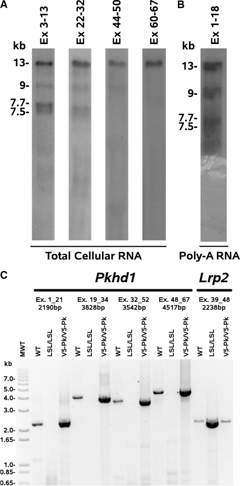
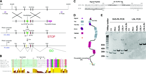
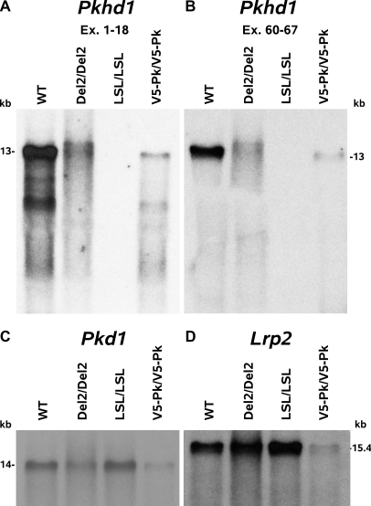
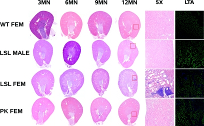
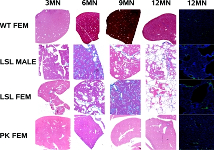
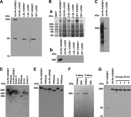
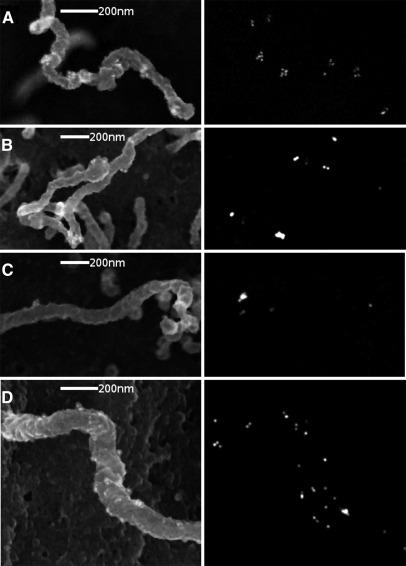
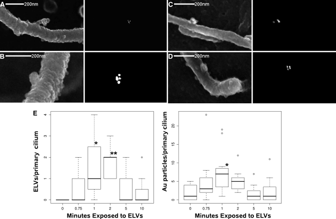
Mutations in the PKHD1 gene, which encodes fibrocystin, cause autosomal recessive polycystic kidney disease (ARPKD). Unfortunately, the lack of specific antibodies to the mouse protein impairs the study of splicing, post-translational processing, shedding, and temporal and spatial expression of endogenous fibrocystin at the cellular and subcellular level. Here, we report using a knock-in strategy to generate a null Pkhd1 strain and a strain that expresses fibrocystin along with two SV5-Pk epitope tags engineered in-frame into the third exon, immediately C-terminal to the signal-peptide cleavage site in a poorly conserved region. By 6 mo of age, the Pkhd1-null mouse develops massive cystic hepatomegaly and proximal tubule dilation, whereas the mouse with epitope-tagged fibrocystin has histologically normal liver and kidneys at 14 mo. Although Pkhd1 was believed to generate many splice forms, our western analysis resolved fibrocystin as a 500 kD product without other forms in the 15-550 kD range. Western analysis also revealed that exosome-like vesicles (ELVs) secrete the bulk of fibrocystin in its mature cleaved form, and scanning electron microscopy identified that fibrocystin on ELVs attached to cilia. Furthermore, the addition of ELVs with epitope-tagged fibrocystin to wild-type cells showed that label transferred to primary cilia within 5 min. In summary, tagging of the endogenous Pkhd1 gene facilitates the study of the glycosylation, proteolytic cleavage, and shedding of fibrocystin.


Figures

Figure 1.
Figure 1.
Pkhd1 shows minimal differential splicing. Northern blotting and RT–PCR of mouse kidney mRNA for Pkhd1 and Lrp2 (megalin): 0.5% agarose, MOPS formaldehyde northern blots, (A) Mouse kidney total cellular RNA probed with probes to mouse Pkhd1 exons 1–13, 22–32, 44–50 and 60–67. The full length product is 13kb, whereas the smaller forms are at 9, 7.7 and 7.5kb. (B) Mouse kidney poly–A mRNA probed with a probe to mouse Pkhd1 exons 1–18, showing that the Pkhd1 and its smaller forms are poly-adenylated. (C) Reverse transcription PCR on poly–A selected mRNA from WT, Pkhd1del2/del2, Pkhd1LSL(−)/LSL(−) and Pkhd1Pk(+)/Pk(+) kidney. PCRs were performed from exons 1 to 21, 19 to 34, 32 to 52 and 48 to 67 and generated species with the expected molecular mass when compared with the mouse sequence of fibrocystin [gi 126157465 ref NM 153179.3 ]. As a loading control we amplified Lrp2 to show that an excess of Pkhd1LSL(−)/LSL(−) mRNA was used in these assays and that the Pkhd1 in the Pkhd1LSL(−)/LSL(−) mouse is completely transcriptionally silent.
Figure 2.
Figure 2.
The strategy used to produce the Pkhd1LSL(−)/LSL(−) and Pkhd1Pk(+)/Pk(+) mouse. (A) A loxP flanked puromycin N–acetyltransferase (pac) and SV–40 derived transcriptional STOP cassette was inserted into Intron–2 of the Pkhd1, LSL. This terminated all Pkhd1 transcripts after this point. Two SV5–Pk epitope tags where also inserted in frame into exon–3 of the Pkhd1 gene. (B) A T–Coffee alignment of the signal peptides and first 27 amino acids of five vertebrate fibrocystins. The insertion site for the tag was chosen so that it was in a region of poor overall conservation and at the extreme N–terminus of the mature protein (once the signal peptide is removed in the endoplasmic reticulum). (C) The protein sequence of the signal peptide, SV5–Pk tag and mature fibrocystin junction in the Pkhd1Pk(+)/Pk(+) mouse. (D) Schematic diagram of the fibrocystin protein showing the SV5–Pk tag site and the pro–protein convertase site. (E) Diagnostic PCRs for the detection of the Pkhd1Pk(+) and Pkhd1LSL(−) alleles (Concise Methods).
Figure 3.
Figure 3.
Pkhd1LSL(−)/LSL(−) mice are null for Pkhd1 mRNA whereas Pkhd1Pk(+)/Pk(+) mice have normal levels of Pkhd1 mRNA. Northern blots for Pkhd1, Pkd1 and Lrp2 (megalin): Northern blots of total mouse kidney RNA, run on a MOPS, formaldehyde, 0.5% agarose gel. WT, Pkhd1del2/del2, Pkhd1LSL(−)/LSL(−) and Pkhd1Pk(+)/Pk(+) kidney RNA probed with (A) the first 18 exons of Pkhd1, (B) the last 7 exons of Pkhd1, (C) a Pkd1 cDNA probe and (D) a Lrp2 megalin probe. The Pkhd1LSL(−)/LSL(−) mouse does not make a Pkhd1 product with either Pkhd1 probe but does synthesize Pkd1 and Lrp2 mRNA. The Pkhd1Pk(+)/Pk(+) kidney lane is somewhat under loaded, but the ratios of Pkhd1 and Lrp2/Pkd1 mRNA are similar to the control. This is reflected in the quantitative PCR in Supplemental Figure 2.
Figure 4.
Figure 4.
Female Pkhd1LSL(−)/LSL(−) mice develop proximal tubule dilatation and cysts, whereas male Pkhd1LSL(−)/LSL(−) mice and both sexes of Pkhd1Pk(+)/Pk(+) mice have cyst-free kidneys. Comparison of WT, Pkhd1Pk(+)/Pk(+) and Pkhd1LSL(−)/LSL(−) kidneys: Comparison of the renal phenotype in WT, male Pkhd1LSL(−)/LSL(−), female Pkhd1LSL(−)/LSL(−) and female Pkhd1Pk(+)/Pk(+) animals. Both Pkhd1LSL(−)/LSL(−) and Pkhd1Pk(+)/Pk(+) animals were inbred to F10 on a C57BL/6J background and compared with C57BL/6J WT mice. Only female Pkhd1LSL(−)/LSL(−) mice begin to develop PT dilation at 3 to 6 mo of age. This is confirmed by immunohistology with Lotus tetragonolobus lectin (LTL) which is specific for the PT (DAPI counter-stain). The female Pkhd1Pk(+)/Pk(+) mice have normal kidneys at 12 mo of age.
Figure 5.
Figure 5.
Both sexes of Pkhd1LSL(−)/LSL(−) mice develop polycystic liver disease and fibrosis, whereas Pkhd1Pk(+)/Pk(+) mice always have normal livers. Comparison of WT, Pkhd1Pk(+)/Pk(+) and Pkhd1LSL(−)/LSL(−) liver: Comparison of the liver phenotype in WT, male Pkhd1LSL(−)/LSL(−), female Pkhd1LSL(−)/LSL(−) and female Pkhd1Pk(+)/Pk(+) animals. Both Pkhd1LSL(−)/LSL(−) and Pkhd1Pk(+)/Pk(+) animals were inbred to F10 on a C57BL/6J background and compared with C57BL/6J WT mice. Both male and female Pkhd1LSL(−)/LSL(−) mice develop liver cysts and fibrosis at 3 mo of age and this worsens with age until the liver is completely replaced by cysts at 12 mo of age. This is confirmed by staining with PCK–26 (IgG1 monoclonal antibody ab6401) anti–cytokeratins 5, 6, and 8, which shows that the cyst lining are epithelial in origin (DAPI counter-stain). Both sexes of the Pkhd1Pk(+)/Pk(+) mouse have normal livers at 12 mo and this has been confirmed to the 14 mo time point.
Figure 6.
Figure 6.
SV5-Pk tagged fibrocystin is easily detected in the kidney, urine, and urinary ELVs. (A). A) 4–12% MOPS PAGE gel resolving from 15–550 kD, there is a 500 kD band in Pkhd1Pk(+)/Pk(+) kidney membrane preparation but no tagged material was seen in Pkhd1LSL(−)/LSL(−) or WT tissues. Note: there is an endogenous band at 50 kD which cross reacts with SV5–Pk1 antibody (this can be used as a loading control). (B) (a) Coomassie blue staining of a duplicate 4 to 12% MOPS PAGE gel in A; (b) 4% slab gel showing that fibrocystin runs as a doublet. (C) 3–8% Nu–PAGE gel comparing 30 μg of Pkhd1Pk(+)/Pk(+) kidney and liver membrane protein, probed with SV5-Pk1 and a HRP conjugated secondary antibody, the liver contains a small fraction of the kidney fibrocystin as biliary epithelial cells make up about 1% of hepatic cells. (D) 3–8% Nu–PAGE gel. Kidney membrane preparations were treated with PNGase and Endo H. PNGase results in fibrocystin resolving at 450 kD, whereas Endo H causes approximately half the kidney fibrocystin to resolve at 450 kD implying that about half of the fibrocystin is in the ER. In the case of Pkhd1Pk(+)/Pk(+) ELVs, fibrocystin shifts from 450 kD to 390 kD after treatment with PNGase (implying that it has undergone the pro–protein convertase cleavage event upon release from the cell). Upon treatment with Endo H there is a very slight decrease in size, c10 kD, implying that the bulk of the glycosylation is mature complex type carbohydrate (post–Golgi). (E) Distribution of fibrocystin in urine of the Pkhd1Pk(+)/Pk(+) mouse. We can use the SV5–Pk1 antibody to detect fibrocystin in unconcentrated urine and again we detected no smaller forms. (F) 200μl of fresh Pkhd1Pk(+)/Pk(+) mouse urine was harvested and 100μl brought to 1x Complete proteinase. This 100μl was centrifuged at 100,000g for 1 h at 4 °C and the supernatant carefully removed and the pellet resuspended. The remaining 100μl was then incubated at 37 °C for 6 h without proteinase inhibitors and the pellet and supernatant collected. A 3–8% MOPS western showed that all of the fibrocystin remained in the PKD–ELV fraction despite the prolonged incubation at 37 °C, showing that the cleaved fibrocystin is firmly attached to the PKD–ELVs. (G) To exclude the possibility of differential splice forms or other processed forms we ran four different ELV samples on a 4 to 12% gel.
Figure 7.
Figure 7.
Fibrocystin is present on ELVs attached to the primary cilium. Immunolocalization of SV5–Pk tagged fibrocystin in cultured PT cells by ISEM: Primary Pkhd1Pk(+)/Pk(+) PT cells, the affected cell type in Pkhd1LSL(−)/LSL(−) mice, were cultured for 7 d and serum starved for 2 d, fixed in paraformaldehyde/glutaraldehyde and stained for SV5–Pk1 with 15nm protein A gold. (Left) Secondary electrons, (Right) Backscattered electrons (gold). (A–D) primary cilia with adherent PKD–ELVs staining with 15nm gold, there is also gold on the shaft of the primary cilium in D.
Figure 8.
Figure 8.
PKD-ELVs interact with primary cilia and are cleared within 10 minutes. PKD–ELV/primary cilium interaction visualized by SEM: Primary WT PT cells are incubated with PKD–ELVs from Pkhd1Pk(+)/Pk(+) mouse urine. (A and B) WT PT primary cilia at 1 min with (A) a ciliary side branch interacting with SV5-Pk positive PKD–ELVs. (C and D) SV–Pk5 positive PKD–ELVs interacting with a primary cilia at 2 min. Left hand panels secondary electrons, right hand panels backscattered electrons – gold (SV5–Pk label, 15nm gold). (E) The number of exosomes positive for SV5–Pk gold adherent to WT primary cilia and the total amount of gold particles on primary cilium after adding 1:10 dilution of Pkhd1Pk(+)/Pk(+) urine, as a function of time. For adherent PKD–ELVs the time points 1 and 2 min were significant versus control (T = 0) at P = 0.0015, and P = 0.001 respectively (an ELV is defined as a 100nm vesicle attached to the primary cilium with >1 gold particle), for total gold per primary cilium the 1 min time point was significant versus control P = 0.038 (One–way ANOVA with Tukey's HSD, data presented as Tukey box plots).

Comment in

  • Tagged fibrocystin sheds its secrets.
    Patel V. Patel V. J Am Soc Nephrol. 2011 Dec;22(12):2148-50. doi: 10.1681/ASN.2011101005. Epub 2011 Nov 11. J Am Soc Nephrol. 2011. PMID: 22080423 Free PMC article. No abstract available.

References

    1. Zerres K, Mucher G, Becker J, Steinkamm C, Rudnik-Schoneborn S, Heikkila P, Rapola J, Salonen R, Germino GG, Onuchic L, Somlo S, Avner ED, Harman LA, Stockwin JM, Guay-Woodford LM: Prenatal diagnosis of autosomal recessive polycystic kidney disease (ARPKD): Molecular genetics, clinical experience, and fetal morphology. Am J Med Genet 76: 137–144 1998 - PubMed
    1. Jorgensen MJ: The ductal plate malformation. Acta Pathol Microbiol Scand Suppl: 1–87, 1977 - PubMed
    1. Zerres K, Rudnik-Schoneborn S, Steinkamm C, Becker J, Mucher G: Autosomal recessive poly-cystic kidney disease. J Mol Med 76: 303–309, 1998 - PubMed
    1. Ward CJ, Hogan MC, Rossetti S, Walker D, Sneddon T, Wang X, Kubly V, Cunningham JM, Bacallao R, Ishibashi M, Milliner DS, Torres VE, Harris PC: The gene mutated in autosomal recessive polycystic kidney disease encodes a large, receptor-like protein. Nat Genet 30: 259–269, 2002 - PubMed
    1. Kaimori JY, Nagasawa Y, Menezes LF, Garcia-Gonzalez MA, Deng J, Imai E, Onuchic LF, Guay-Woodford LM, Germino GG: Polyductin undergoes notch-like processing and regulated release from primary cilia. Hum Mol Genet 16: 942–956, 2007 - PMC - PubMed

Publication types