. 2011 May;44(2):337-47.
doi: 10.4103/0970-0358.85355.

Malignant tumours of the hand and wrist

Affiliations

Malignant tumours of the hand and wrist

Binu P Thomas et al. Indian J Plast Surg. 2011 May.

Abstract

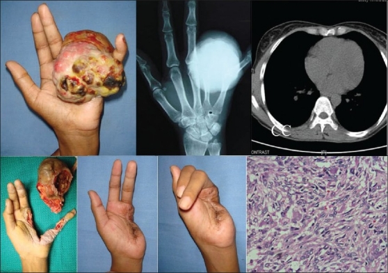
Malignant tumours are rare in the hand and wrist. The clinical presentation may be similar to that of a benign lesion and a high index of suspicion is necessary so that such lesions are not missed by the treating surgeon. Out of a total of 657 tumours/tumour-like lesions of the hand and wrist seen in a tertiary referral centre in a 10-year period, a total of 39 tumours were identified as malignant (5.9%) and of which majority had origin from the skin (53.8%). The management of these tumours is primarily surgical. Limb salvage surgery may be applied when appropriate, though eradication of disease should be the primary goal rather than preservation of function. A multimodal approach is necessary for appropriate management including chemotherapy and radiotherapy.

Keywords: Hand tumours; malignant hand tumours; malignant tumour Rev.


Conflict of interest statement

Conflict of Interest: None declared.

Figures

Diagram 1
Diagram 1
Line drawing to denote levels of amputation for tumours occurring at various sites
Figure 1
Figure 1
Squamous cell carcinoma (SCC) (a) Infiltrating SCC left wrist, arsenical keratosis both hands. (b) Amputation left hand. (c) SCC right palm. (d) Excision and PPIA flap. (e) Hand E stain ×100: nests of malignant squamous epithelial cells with keratin pearls
Figure 2
Figure 2
Squamous cell carcinoma (a) Squamous cell carcinoma from burn scar (Marjolins ulcer). (b) Subungual squamous cell carcinoma. (c,d,e) 20 year history of ulcer in the palm, four previous surgeries elsewhere, moderately differentiated SCC. Limited ray excision, epitrochlear lymph node removal, axillary clearance and local radiotherapy, three cycles of 60 Gy and chemotherapy with paclitaxel and carboplatin six cycles given
Figure 3
Figure 3
Malignant melanoma (MM), (a) MM thumb, (b) Axillary metastasis, (c) High power(400×) view: dark brown granular melanin pigment within the tumour cells
Figure 4
Figure 4
Dermatofibrosarcoma protuberans (DFSP), (a,b) DFSP recurrence. Ray resection done. (c) Trucut biopsy specimen: characteristic diffuse CD34 cytoplasmic positivity on immunohistochemistry (d)
Figure 5
Figure 5
Synoviosarcoma, (a) swelling right thumb base, (b) B1, B2: X-ray and MRI showing the lesion with involvement of tendon sheaths and vascular structures, (c,d) A radical resection was done. (e) ×200, Hand E staining showing sheets and nests of polygonal and spindle cells. (f,g) Epithelial membrane antigen EMA and CD99 positivity are shown
Figure 6
Figure 6
(a) Fibrosarcoma (FS), (a,b,c) Swelling 1st web space with MRI showing a well-defined rounded mass reported as schwannoma. Wide excision following needle biopsy reported as low-grade FS, (b) Epitheloid sarcoma (ES)
Figure 7
Figure 7
Extraskeletal myxoid chondrosarcoma (ESMCS), (a,b) 24-year-old lady presented with mass, index finger, (c) X-ray: Expansile soft tissue mass with some lysis of the proximal phalanx, (d) Biopsy ESMCS underwent a ray resection, (e,f) Excellent function and cosmesis and is still being followed up after four years
Figure 8
Figure 8
Malignant spindle cell tumour (MSCT), (a,b) Five-month-old baby with four months history of swelling in the thumb, biopsy showed a MSCT with D/d infantile fibrosarcoma/rhabdomyosarcoma/leiomyosarcoma. Child underwent below elbow amputation. Post op chemotherapy with vincristine and D-actinomycin. No further follow up and status unknown. (c,d) Hand E showing sheets and fascicles of spindle-shaped cells with focal herring bone pattern. This spindle cell sarcoma shows focal positivity for desmin and smooth muscle actin (SMA)
Figure 9
Figure 9
Ewings sarcoma/PNET
Figure 10
Figure 10
Ewing's sarcoma/PNET, (a,b) 17-year-old boy with swelling and pain of the middle finger of one year, (c) X-rays: Expansile, sclerotic lesion of P1, (d) Ray excision followed by six cycles of vincristine, actinomycin, cyclophosphamide, (e,f) Follow up bone scans revealed a lytic lesion in the 4th rib for which local radiotherapy was given. He remains disease free on a three-year follow up, (g) Hand E staining showing lobules of small round cells, with hyperchromatic nuclei and scant cytoplasm exhibiting increased mitotic activity, (h) Immunohistochemistry shows tumour cells are CD99 positive
Figure 11
Figure 11
Giant cell tumour with cellular atypia, GCT recurrence
Figure 12
Figure 12
Giant cell tumour recurrence
Figure 13
Figure 13
Secondaries in the hand, (a,b) X ray shows metastasis to thumb, (c,d) Patient with T cell lymphoma with cutaneous infiltration

References

    1. Mankin HJ, Lange TA, Spanier SS. The hazards of biopsy in patients with malignant primary bone and soft-tissue tumors. J Bone Joint Surg Am. 1982;64:1121–7. - PubMed
    1. Enneking WF, Spanier SS, Goodman MA. A system for the surgical staging of musculoskeletal sarcoma. Clin Orthop Relat Res. 1980;153:106–20. - PubMed
    1. Green FL, Page DL, Fleming ID, Fritz AG, Balch CM, Haller G, et al. 6th edition. New York: Springer-Verlag; 2002. AJCC cancer staging handbook; pp. 213–5.
    1. Wunder J. Musculoskeletal tumors: staging systems., in Orthopaedic knowledge update: musculoskeletal tumors. In: Menendez LR, editor. Rosemont: 2002. pp. 21–7.
    1. Smith RJ. Tumors of the hand: who is best qualified to treat tumors of the hand? J Hand Surg Am. 1977;2:251–2. - PubMed