. 2011 Jul;2(3):141-52.
doi: 10.4103/0975-3583.85260.

An overview of lymphatic vessels and their emerging role in cardiovascular disease

Affiliations

An overview of lymphatic vessels and their emerging role in cardiovascular disease

Dennis Jones et al. J Cardiovasc Dis Res. 2011 Jul.

Abstract

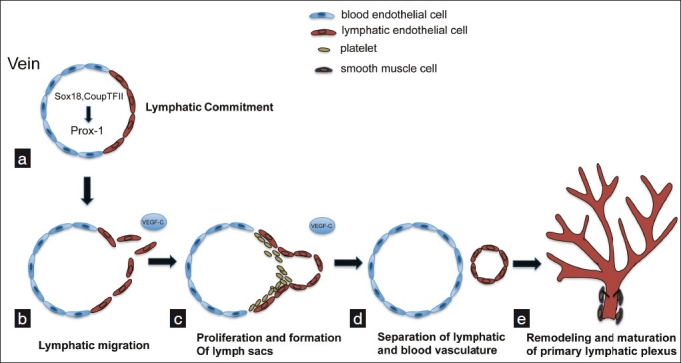
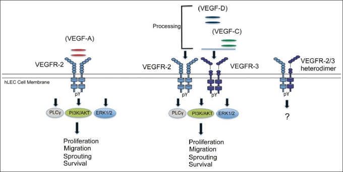
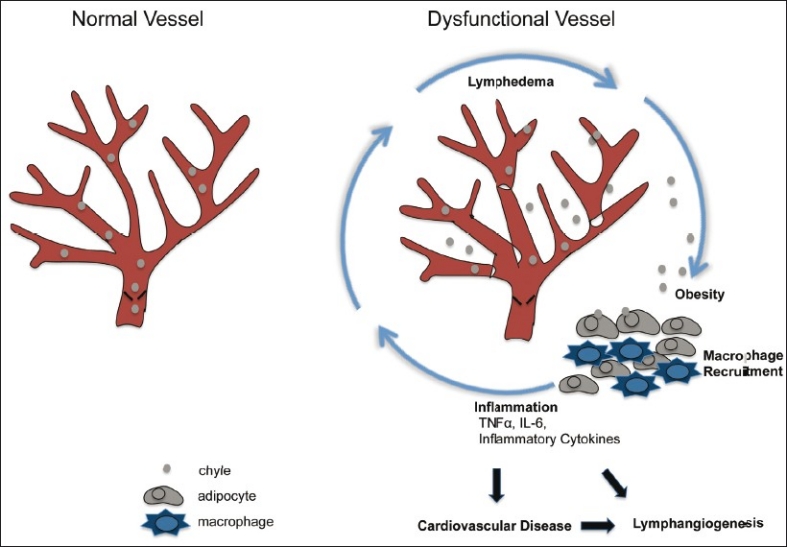
Over the past decade, molecular details of lymphatic vessels (lymphatics) have been rapidly acquired due to the identification of lymphatic endothelial-specific markers. Separate from the cardiovascular system, the lymphatic system is also an elaborate network of vessels that are important in normal physiology. Lymphatic vessels have the unique task to regulate fluid homeostasis, assist in immune surveillance, and transport dietary lipids. However, dysfunctional lymphatic vessels can cause pathology, while normal lymphatics can exacerbate pathology. This review summarizes the development and growth of lymphatic vessels in addition to highlighting their critical roles in physiology and pathology. Also, we discuss recent work that suggests a connection between lymphatic dysfunction and cardiovascular disease.

Keywords: Cardiovascular disease; inflammation; lymphatic vessels; lymphedema; vegf receptor-3.


Conflict of interest statement

Conflict of Interest: None declared.

Figures

Figure 1
Figure 1
Development of the murine lymphatic vasculature. (a) Lymphatic competent “precursors” begin to express Sox-18 and CoupTFII, which subsequently induce expression of Prox-1, which is responsible for induction of and maintenance of the LEC phenotype. (b) A mesenchymal source of VEGF-C initiates sprouting of LECs and formation of primitive lymph sacs, through VEGFR-3. With the assistance of platelets and a continued source of VEGF-C, lymphatic cells separate from veins and proliferate to form a primitive lymphatic network (c, d), which is later remodeled into the mature lymphatic vascular system that features organization into capillaries and larger collecting vessels (e)
Figure 2
Figure 2
VEGF-VEGFR signaling in lymphangiogenesis. Of the VEGFR family proteins, VEGFR-2 and VEGFR-3 are strongly expressed in hLECs in vivo and exist as homodimers and heterodimers. VEGF-A activates VEGFR-2, whereas VEGF-C and VEGF-D activate both VEGFR-2 (after proteolytic cleavage) and VEGFR-3. In human lymphatic EC, VEGF-A preferentially induces phosphorylation of PLC-γ. In contrast, VEGF-C and D preferentially activate the Akt pathway. Both growth factors strongly activate the MAPK pathway. These signaling pathways have been shown to lead to effector functions of LECs, namely migration, sprouting, proliferation, enlargement (growth), survival and tube formation in vivo and in vivo. pY, tyrosine phosphorylation
Figure 3
Figure 3
Proposed working model for dysfunctional lymphatic vessel contribution to cardiovascular disease (evidence from literature in text). Persons diagnosed with metabolic syndrome are at greater risk to develop cardiovascular disease. The metabolic syndrome is a condition diagnosed with the appearance of several risk factors in an individual, and obesity is considered a major risk factor. Dysfunctional lymphatic vessels are unable to properly drain lymph and chyle (shown here). This can cause edema and increased fluid volume leading to chylous ascites and chylothorax. Furthermore, chyle (including chylomicrons) can leak from compromised lymphatics and influence surrounding reservoirs of fat. Lymph can cause maturation of preadipocytes and growth of adipose tissue. Secretion of inflammatory cytokines from adipose tissue and macrophages is exacerbated in obese individuals. Recruitment of macrophages to adipose tissue is increased and excess inflammation can increase lymphangiogenesis and add further detriment to lymphatic vessels, amplifying the cycle, and may eventually contribute to cardiovascular disease

References

    1. Aselli G, Bassano C, Falcini D, Settala S, Tadino A. Mediolani: Apud Jo. Baptistam Bidellium; 1627. De lactibus, sive lacteis venis, quarto vasorum mesaraicorum genere, novo invento.Dissertatio, qua sententiae anatomicae multae, vel perperam receptae convelluntur vel parum perceptae illustrantur.
    1. Wigle JT, Oliver G. Prox1 function is required for the development of the murine lymphatic system. Cell. 1999;98:769–78. - PubMed
    1. Breiteneder-Geleff S, Soleiman A, Horvat R, Amann G, Kowalski H, Kerjaschki D. Podoplanin--a specific marker for lymphatic endothelium expressed in angiosarcoma. Verh Dtsch Ges Pathol. 1999;83:270–5. - PubMed
    1. Banerji S, Ni J, Wang SX, Clasper S, Su J, Tammi R, et al. Lyve-1, a new homologue of the cd44 glycoprotein, is a lymph-specific receptor for hyaluronan. J Cell Biol. 1999;144:789–801. - PMC - PubMed
    1. Sabin F. On the origin of the lymphatic system from the veins, and the development of the lymph hearts and thoracic duct in the pig. Am J Anat. 1902;1:367–89.