Review
. 2010;10(10):9002-25.
doi: 10.3390/s101009002.

Glucose sensing neurons in the ventromedial hypothalamus

Affiliations
Review

Glucose sensing neurons in the ventromedial hypothalamus

Vanessa H Routh. Sensors (Basel). 2010.

Abstract

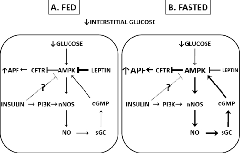
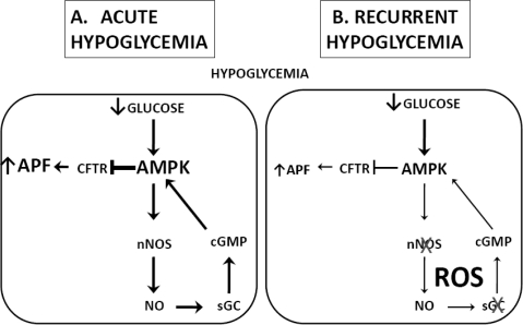
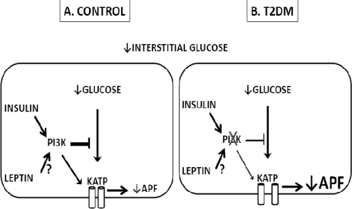
Neurons whose activity is regulated by glucose are found in a number of brain regions. Glucose-excited (GE) neurons increase while glucose-inhibited (GI) neurons decrease their action potential frequency as interstitial brain glucose levels increase. We hypothesize that these neurons evolved to sense and respond to severe energy deficit (e.g., fasting) that threatens the brains glucose supply. During modern times, they are also important for the restoration of blood glucose levels following insulin-induced hypoglycemia. Our data suggest that impaired glucose sensing by hypothalamic glucose sensing neurons may contribute to the syndrome known as hypoglycemia-associated autonomic failure in which the mechanisms which restore euglycemia following hypoglycemia become impaired. On the other hand, increased responses of glucose sensing neurons to glucose deficit may play a role in the development of Type 2 Diabetes Mellitus and obesity. This review will discuss the mechanisms by which glucose sensing neurons sense changes in interstitial glucose and explore the roles of these specialized glucose sensors in glucose and energy homeostasis.

Keywords: diabetes; fasting; glucose-excited neurons; glucose-inhibited neurons; hypoglycemia; hypoglycemia-associated autonomic failure; insulin; leptin; obesity.


Figures

Figure 1.
Figure 1.
Regulation of the glucose sensitivity of NPY-GI neurons in fed and fasted conditions. Decreased interstitial glucose levels are translated into decreased intracellular glucose levels resulting in an increased AMP/ATP ratio. Increased AMP/ATP activates AMPK which enhances NO-sGC-cGMP signaling. Increased levels of cGMP are needed for full AMPK activation and closure of the CFTR chloride conductance in response to decreased glucose. CFTR closure leads to depolarization and increased action potential frequency. In the fed state, elevated leptin levels cause tonic inhibition of AMPK and attenuation of the response of GI neurons to decreased glucose. Insulin levels are also elevated in the fed state. Insulin increases nNOS activation via PI3K which could lead to enhanced responses to decreased glucose. It is also possible that insulin may inhibit AMPK in GI neurons since this effect of insulin has been demonstrated in hypothalamic tissue. AMPK inhibition would contribute to a low sensitivity to glucose decreases. However, the effects of insulin on the glucose sensitivity of GI neurons have not yet been evaluated. In the fasted state, both leptin and insulin levels are reduced. The reduction in leptin-induced AMPK inhibition leads to an enhanced response of GI neurons to decreased glucose. Whether insulin contributes to or opposes this change in glucose sensitivity is not known. Abbreviations: NPY (neuropeptide Y), GI (glucose-inhibited), AMPK (AMP-activated protein kinase), sGC (soluble guanylyl cyclase), cGMP (cyclic GMP), CFTR (cystic fibrosis transmembrane regulator), PI3K (phosphatidylinositol-3-kinase).
Figure 2.
Figure 2.
Hypothetical mechanism underlying effects of recurrent hypoglycemia on the glucose sensitivity of VMH GI neurons. Acute hypoglycemia (A) leads to activation of AMPK and the NO-sGC-cGMP signaling pathway. cGMP further activates AMPK leading to closure of the CFTR, depolarization and increased action potential frequency. Hypoglycemia also increases ROS levels. The combination of NO and ROS production may cause S-nitrosylation of nNOS and sGC. S-nitrosylation of these enzymes leads to a decrease in their activity and a reduction in NO signaling. Therefore, recurrent hypoglycemia (B) would lead to decreased sensitivity of GI neurons to reduced glucose. Abbreviations: VMH (ventromedial hypothalamus), GI (glucose-inhibited), AMPK (AMP-activated protein kinase), sGC (soluble guanylyl cyclase), cGMP (cyclic GMP), CFTR (cystic fibrosis transmembrane regulator), ROS (reactive oxygen species).
Figure 3.
Figure 3.
Hormonal Regulation of the glucose sensitivity of VMH GE Neurons. Hormonal regulation of the glucose sensitivity of VMH GE neurons in controls (A) and T2DM (B). Decreased interstitial glucose levels are translated into decreased intracellular glucose. This decreases the ATP/ADP ratio, opens the KATP channel and hyperpolarizes the cell leading to a decrease in action potential frequency. Insulin, via PI3K, attenuated KATP activation in response to decreased glucose. Since PI3K also mediates leptins effects on GE neurons, it is possible that leptin similarly reduces GE responses to decreased glucose however this has not been tested. Moreover, leptin is known to have pleiotropic effects on VMH GE neurons. In T2DM, insulin resistance leads to an increased responsiveness of GE neurons to glucose decreases which is normalized by enhancing PI3K signaling. Whether leptin resistance during T2DM contributes to enhanced responses of GE neurons to decreased glucose remains to be tested. Abbreviations: VMH (ventromedial hypothalamus), GE (glucose-excited), T2DM (type 2 diabetes mellitus), KATP (ATP-sensitive potassium channel), PI3K (phosphatidylinositol-3-kinase).

Comment in

Similar articles

Cited by

References

    1. Schwartz MW, Woods SC, Porte D, Seeley RJ, Baskin DG. Central nervous system control of food intake. Nature. 2000;404:661–671. - PubMed
    1. Niswender KD, Schwartz MW. Insulin and leptin revisited: Adiposity signals with overlapping physiological and intracellular signaling capabilities. Front. Neuroendocrinol. 2003;24:1–10. - PubMed
    1. Shin AC, Pistell PJ, Phifer CB, Berthoud HR. Reversible suppression of food reward behavior by chronic mu-opioid receptor antagonism in the nucleus accumbens. Neuroscience. 2010;170:580–588. - PMC - PubMed
    1. King BM. The rise, fall, and resurrection of the ventromedial hypothalamus in the regulation of feeding behavior and body weight. Physiol. Behav. 2006;87:221–244. - PubMed
    1. Elmquist JK. Hypothalamic pathways underlying the endocrine, autonomic, and behavioral effects of leptin. Physiol. Behav. 2001;74:703–708. - PubMed

Publication types

LinkOut - more resources