. 2011 Oct;7(10):e1002282.
doi: 10.1371/journal.ppat.1002282. Epub 2011 Oct 13.

A wide extent of inter-strain diversity in virulent and vaccine strains of alphaherpesviruses

Affiliations

A wide extent of inter-strain diversity in virulent and vaccine strains of alphaherpesviruses

Moriah L Szpara et al. PLoS Pathog. 2011 Oct.

Abstract

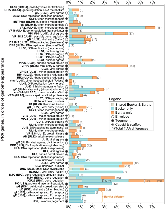
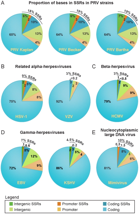
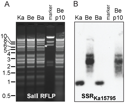
Alphaherpesviruses are widespread in the human population, and include herpes simplex virus 1 (HSV-1) and 2, and varicella zoster virus (VZV). These viral pathogens cause epithelial lesions, and then infect the nervous system to cause lifelong latency, reactivation, and spread. A related veterinary herpesvirus, pseudorabies (PRV), causes similar disease in livestock that result in significant economic losses. Vaccines developed for VZV and PRV serve as useful models for the development of an HSV-1 vaccine. We present full genome sequence comparisons of the PRV vaccine strain Bartha, and two virulent PRV isolates, Kaplan and Becker. These genome sequences were determined by high-throughput sequencing and assembly, and present new insights into the attenuation of a mammalian alphaherpesvirus vaccine strain. We find many previously unknown coding differences between PRV Bartha and the virulent strains, including changes to the fusion proteins gH and gB, and over forty other viral proteins. Inter-strain variation in PRV protein sequences is much closer to levels previously observed for HSV-1 than for the highly stable VZV proteome. Almost 20% of the PRV genome contains tandem short sequence repeats (SSRs), a class of nucleic acids motifs whose length-variation has been associated with changes in DNA binding site efficiency, transcriptional regulation, and protein interactions. We find SSRs throughout the herpesvirus family, and provide the first global characterization of SSRs in viruses, both within and between strains. We find SSR length variation between different isolates of PRV and HSV-1, which may provide a new mechanism for phenotypic variation between strains. Finally, we detected a small number of polymorphic bases within each plaque-purified PRV strain, and we characterize the effect of passage and plaque-purification on these polymorphisms. These data add to growing evidence that even plaque-purified stocks of stable DNA viruses exhibit limited sequence heterogeneity, which likely seeds future strain evolution.


Conflict of interest statement

The authors have declared that no competing interests exist.

Figures

Figure 1
Figure 1. Genome organization of PRV Kaplan and comparison of sequence conservation with strains Becker and Bartha.
A) Open reading frames (ORFs) are plotted spatially along the genome, along with their untranslated regions (UTRs). The genome proceeds from the Unique Long (UL) region into the Unique Short (US) region, with US being flanked by long inverted repeats (IR, TR). The large-latency transcript (LLT) is not translated. A horizontal bar connects the spliced portions of the UL15 ORF; splicing in the 5′ UTR of US1 is not shown for space reasons. B) Graph depicts DNA sequence conservation between PRV Kaplan and PRV Becker or PRV Bartha. Conservation is calculated from a multiple sequence alignment, and the conservation score between any two genomes is plotted from a sliding 100 bp window. C) Short sequence repeats (SSRs) are plotted as they occur along the PRV Kaplan genome. SSRs include minisatellites (repeat unit ≥10 bp), microsatellites (repeat unit <10 bp), and homopolymers (minimum length 6). D) A phylogenetic tree based on a whole-genome multiple sequence alignment demonstrates the closer relationship of PRV strains Kaplan and Bartha. Bootstrap values are shown at branch points (see Methods for details). The same result was obtained using nucleotide sequences of gC (data not shown), as was done for several recent PRV phylogenetic comparisons , , .
Figure 2
Figure 2. BamHI RFLP confirmation of PRV genome assemblies.
A) Location of major BamHI fragments along the PRV Kaplan genome. Fragments are identified by historical fragment numbering . Genome position in kilobase pairs (kb) is listed below the fragments, with the large inverted repeats IR and TR shown as green boxes. B) RFLP analysis of BamHI fragments of PRV strains Kaplan (Ka), Becker (Be), Bartha (Ba), an unpurified Kaplan stock (Ka*np), and a Becker stock passaged 10 times in vitro (Be*p10). Positions of a standard marker are noted on the left. Major BamHI fragments, and their predicted size in each strain, are indicated to the right at their approximate height in the gel image. The Bartha deletion causes a major size shift in BamHI fragment 7 (boxed; now 3.1 kb); the new band co-migrates with BamHI fragment 11 (also 3.1 kb). All bands except the variable fragments 10 and 12 (red asterisks) match their predicted sizes. Arrows from Kaplan through Becker and Bartha columns indicate bands that are predicted to be of equivalent size in all three strains.
Figure 3
Figure 3. Protein coding variation in PRV Bartha and Becker, vs. the reference strain Kaplan.
The percent of AAs differing in PRV Becker and Bartha, vs. the new reference genome of PRV Kaplan, are plotted in order of occurrence along the PRV genome (top to bottom). Eight proteins show no inter-strain variation in coding sequence. The total number of differences (Tables 1– 3) have been normalized to protein length. Protein names and functions are listed on the left, along with a symbol indicating if the protein product is a known virion component. AA differences from the reference strain Kaplan are categorized as being unique to the vaccine strain PRV Bartha (orange), unique to the virulent strain PRV Becker (blue), or shared (observed in both Bartha and Becker; gray). The four proteins affected by the deletion in Bartha's US region are bracketed at the bottom.
Figure 4
Figure 4. Inter-strain variation in protein levels of gH.
A) Western blot analysis of infected cell lysates demonstrates that PRV Bartha produces gH (UL22) comparable to that in virulent strains. PRV Becker displays slightly higher and/or differentially glycosylated levels of gH than the other two strains. Levels of the capsid protein VP5 (UL19) are shown for comparison and as a loading control. B) Ratio of gH vs. VP5 in each sample, using the ImageJ Gel Analyzer module. Equivalent amounts of protein were loaded in each lane. The blot was cut, with VP5 measured on the upper half and gH on the lower half to demonstrate equivalent levels of infection in each lysate. The same lysates were used for the analyses in Figure S3 (in Text S1); these are representative of three separate experiments. The conditions required to visualize the two bands of gH precluded measurement of cellular actin on the same blot. Positions of a standard marker are noted on the left.
Figure 5
Figure 5. Inter-strain diversity in protein coding sequences, in PRV as compared to HSV-1.
The total number of amino acid differences between three strains of PRV (strains Kaplan, Becker, Bartha) is normalized for protein length and plotted with data for the homologous proteins of HSV-1 (strains 17, F, H129) . Blue color highlights variable proteins where inter-strain variation reaches similar levels in PRV and HSV-1, while orange highlights proteins that are much more variable in PRV than HSV-1, and green highlights the converse. Boxed proteins UL31 and UL20 show no variations in these six strains of alphaherpesvirus; UL31 also shows no coding variation across 18 strains of VZV , . Proteins without homologues in both viruses are excluded, as are proteins in the Bartha deletion region. Table S6 lists all protein names, lengths, and percent variation in PRV, HSV-1, and VZV strains.
Figure 6
Figure 6. Prevalence of SSRs in PRV strains and in related DNA viruses.
The proportion of bases in each genome involved in SSRs was calculated for (A) all three PRV strains, as well as for (B) the related human alphaherpesviruses HSV-1 and VZV, (C) the betaherpesvirus HCMV, (D) the gammaherpesviruses EBV and KSHV, and (E) a nucleocytoplasmic large DNA virus, Mimivirus. Pie charts depict what proportion of each genome falls into coding regions, promoter regions (defined as 500 bp upstream of a coding sequence), or open intergenic regions. Exact numbers and types of SSRs per genome are found in Table 4. A complete list of all PRV SSRs is found in Table S7.
Figure 7
Figure 7. Southern blot of CAPRE-estimated SSR lengths.
A) RFLP analysis of SalI fragments of PRV strains Kaplan (Ka), Becker (Be), Bartha (Ba), and a Becker stock passaged 10 times in vitro (Be p10). Positions of one standard marker are noted on the left; another marker lane is shown between the Bartha and Becker p10 lanes (5 bands: 23 kb, 9.4 kb, 6.6 kb, 2.3 kb, 2 kb). Asterisk (*) at 7 kb in the PRV Bartha lane highlights a size shift in the fragment containing the Bartha US-region deletion. B) Southern blot of the same fragments, using a biotinylated probe matching SSRKa17595 (a perfect 15-mer) to reveal the size of the SalI fragment containing this site. Without any SSR content, this fragment would be ∼0.55 kb in PRV Kaplan and Bartha, and ∼0.74 kb in PRV Becker. Based on observed fragment sizes, the 1 kb Kaplan and Bartha fragments each have ∼30 copies of this SSR, while the 2.5 kb average Becker fragment (range 1.6–3.5 kb) has on average ∼120 copies (range 58–184 copies). The variable fragment size in PRV Becker shifts upon passage in vitro (Be p10).
Figure 8
Figure 8. Percent of data supporting polymorphic base calls in PRV genomes.
A) A limited number of polymorphic bases were detected in the plaque-purified strains Kaplan, Becker, and Bartha; these were analyzed to deduce the percent of sequence data supporting the primary vs. alternative base calls. The majority of polymorphic sites show 1–20% support for the alternative base call, or 99–80% support for the primary base call. B) After passaging the purified Becker stock multiple times in vitro (Becker p10), there was no increase in the overall number of polymorphic bases, and only a slight shift in the degree of support for alternative base calls. C) However in an unpurified historical stock of PRV Kaplan, which is the parent of the plaque-purified stock used for sequencing, hundreds of polymorphic bases were observed. Despite the larger quantity of polymorphic bases, the degree of support for the alternative base is similar to that found in the plaque-purified strains. D) Graph displays the alternative base calls for the four most variant polymorphisms in PRV Becker (41–50% bin), and how these specific bases were called in the Becker p10 progeny stock (the height of each base letter corresponds to its frequency). These sites display shifts in base frequency in the Becker p10 stock.

Similar articles

Cited by

References

    1. Roizman B, Pellett PE. The family Herpesviridae: A brief introduction. In: Knipe DM, Howley PM, editors. Fields Virology 4 ed. Philadelphia: Lippincott Williams & Wilkins; 2001. pp. 2381–2397.
    1. Steiner I, Kennedy PG, Pachner AR. The neurotropic herpes viruses: herpes simplex and varicella-zoster. Lancet Neurol. 2007;6:1015–1028. - PubMed
    1. Arvin AM. Varicella-zoster virus. Clin Microbiol Rev. 1996;9:361–381. - PMC - PubMed
    1. Pomeranz LE, Reynolds AE, Hengartner CJ. Molecular biology of pseudorabies virus: impact on neurovirology and veterinary medicine. Microbiol Mol Biol Rev. 2005;69:462–500. - PMC - PubMed
    1. Mettenleiter TC, Keil GM, Fuchs W. Molecular Biology of Animal Herpesviruses. In: Mettenleiter TC, Sobrino F, editors. Animal viruses: molecular biology. xii. Norfolk, UK: Caister Academic Press; 2008. p. 531.

Publication types

MeSH terms

Associated data