. 2011;6(10):e25761.
doi: 10.1371/journal.pone.0025761. Epub 2011 Oct 13.

Production of embryonic and fetal-like red blood cells from human induced pluripotent stem cells

Affiliations

Production of embryonic and fetal-like red blood cells from human induced pluripotent stem cells

Chan-Jung Chang et al. PLoS One. 2011.

Abstract

We have previously shown that human embryonic stem cells can be differentiated into embryonic and fetal type of red blood cells that sequentially express three types of hemoglobins recapitulating early human erythropoiesis. We report here that we have produced iPS from three somatic cell types: adult skin fibroblasts as well as embryonic and fetal mesenchymal stem cells. We show that regardless of the age of the donor cells, the iPS produced are fully reprogrammed into a pluripotent state that is undistinguishable from that of hESCs by low and high-throughput expression and detailed analysis of globin expression patterns by HPLC. This suggests that reprogramming with the four original Yamanaka pluripotency factors leads to complete erasure of all functionally important epigenetic marks associated with erythroid differentiation regardless of the age or the tissue type of the donor cells, at least as detected in these assays. The ability to produce large number of erythroid cells with embryonic and fetal-like characteristics is likely to have many translational applications.


Conflict of interest statement

Competing Interests: The authors have declared that no competing interests exist.

Figures

Figure 1
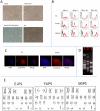
Figure 1. iPS characterization.
A: Phase contrast micrograph illustrating the morphology of human iPS cells on Matrigel. B: FACS analysis. Histograms illustrating expression of surface marker, SSEA-3, SSEA-4, Tra-1-60, and Tra-1-8 on ES and iPS cells. The black histograms represent the IgG control. C: immuno-micrographs illustrating expression of transcription factor Oct3/4. Cells were permeabilized and stained with PE-labeled anti-Oct4 antibodies and with DAPI. Most cells in the iPS and the H1 colonies exhibited nuclear Oct3/4 expression D. LM-PCR analysis. Photograph of a 1% agarose gel illustrating the result of an LM-PCR determination of integration site patterns. E: Karyotyping of iPS derived from different cell of origins.
Figure 2
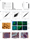
Figure 2. Expression and differentiation analysis.
A: Expression of the virally-transduced reprogramming factors cDNA. Y-axis represent absolute number of copies of cDNA per ng of RNA determined using a standard curved based on a purified PCR product. Expression of the transduced cDNA was mostly silent except for FL iPS C (see text). B: Global expression patterns: Scatter-plot illustrating an Affymetrix micro-array analysis of iPS as compared to H1 and to parental somatic cells. Micro-array results were normalized by RMA. Average of two biological replicates is plotted. Left scatter-plot illustrates expression of H1 and H9 cells, middle scatter-plot, expression of iPSA versus p51R parental cells and right scatter plot expression of iPSA versus H1. iPS are undistinguishable from ES cells in this assay. All iPS were at least at passage 20 when the RNA were extracted. C: Embryoid bodies. Phase contrast micrographs illustrating 5 days EB formed from iPS. D: Spontaneous differentiation: Immuno-fluorescence micrographs illustrating iPS differentiation into the three germ layers. Two days EB were transferred to chamber slides containing ES medium without bFGF. After 7 days, the differentiated cells were stained with FITC labeled antibodies against β-III-tubulin (ectoderm), α-smooth muscle actin (mesoderm) and α-feto-protein (endoderm) and counter-stained with DAPI. Results from 2 different iPS are shown. (Bar = 100μm). E: Micrographs illustrating H and E stained slide of teratomas generated from an iPS. iPS can differentiate into teratomas containing the three germ layers (see also Figure S5).
Figure 3
Figure 3. Hematopoietic differentiation.
A: Production of CD34 positive cells: hES and iPS cells were co-co-cultured for 7 to 35 days with FHB-hTERT. Percentage of cell expressing CD34 was then assessed by FACS using FITC-labeled anti-CD34 antibodies. Most iPS produced similar level of CD34+ cells as H1 except FL iPSC which produced 10 to 15 times more CD34+ cells. B: Giemsa staining of erythroid cells obtained by differentiation of H1 or iPS-derived CD34+ cells obtained from co-cultures with FHB-hTERT for 35 days. After the co-culture, the cells were placed in liquid culture for 14 days (first row) or 21 days (rows 2 and 3) and cytospins were performed. iPS-derived cells have morphologies very similar to ES-derived cells regardless of the age of the donor. C: Chromatograms illustrating HPLC analyses of globins produced by ES or iPS derived erythrocytes. Top left chromatogram: Erythrocytes produced after 14 days of FHB-hTERT co-culture and 14 days of liquid culture express mostly Hb Gower I (ε2ζ2), bottom left: after 10 more days of liquid culture (24D), erythroblast down-regulated ζ-globin leading to the production of Hb Gower 2 (ε2ζ2). Erythroblasts produced after 35 days of co-culture with FH-B-hTERT and 14 days (top right) or 24 days (bottom right) of liquid culture express mostly Hb F (γ2α2). D: Histograms summarizing HPLC results from 9 iPS clones (left) or from triplicated differentiation experiments of H1 ES cells (right). Erythroid cells generated by co-culture of iPS on FhB-hTERT for 14 days exhibit a major ζ to α globins switching during their maturation from basophilic to orthochromatic erythroblasts in liquid culture. Cell generated after 35 days of co-culture on FhB-hTERT express primarily gamma globins and have therefore switched their expression of the β-like globin genes from ε to γ globin as compared to cells obtained after 14 days of co-culture. Globin expression patterns of iPS-derived cells are very similar to what is observed with hESC. E: Flow cytometry data showing that H1 and SK-iPS after co-culture with FHB-hTERT cells for 14 days cells differentiate into cells that express CD34 and CD43. By contrast, FL-iPS C-derived cells, express CD34 but not CD43. After liquid culture ES and iPS-derived cells differentiate into cells that co-express CD34+ and CD45+. FL-iPS C do not proliferate.

References

    1. Takahashi K, Yamanaka S. Induction of pluripotent stem cells from mouse embryonic and adult fibroblast cultures by defined factors. Cell. 2006;126:663–676. - PubMed
    1. Takahashi K, Tanabe K, Ohnuki M, Narita M, Ichisaka T, et al. Induction of pluripotent stem cells from adult human fibroblasts by defined factors. Cell. 2007;131:861–872. - PubMed
    1. Yu J, Vodyanik MA, Smuga-Otto K, Antosiewicz-Bourget J, Frane JL, et al. Induced pluripotent stem cell lines derived from human somatic cells. Science. 2007;318:1917–1920. - PubMed
    1. Wernig M, Meissner A, Foreman R, Brambrink T, Ku M, et al. In vitro reprogramming of fibroblasts into a pluripotent ES-cell-like state. Nature. 2007;448:318–324. - PubMed
    1. Park IH, Zhao R, West JA, Yabuuchi A, Huo H, et al. Reprogramming of human somatic cells to pluripotency with defined factors. Nature. 2008;451:141–146. - PubMed

Publication types

Associated data