. 2011;6(10):e25770.
doi: 10.1371/journal.pone.0025770. Epub 2011 Oct 12.

Age-related impairment of ultrasonic vocalization in Tau.P301L mice: possible implication for progressive language disorders

Affiliations

Age-related impairment of ultrasonic vocalization in Tau.P301L mice: possible implication for progressive language disorders

Clément Menuet et al. PLoS One. 2011.

Abstract

Background: Tauopathies, including Alzheimer's Disease, are the most frequent neurodegenerative diseases in elderly people and cause various cognitive, behavioural and motor defects, but also progressive language disorders. For communication and social interactions, mice produce ultrasonic vocalization (USV) via expiratory airflow through the larynx. We examined USV of Tau.P301L mice, a mouse model for tauopathy expressing human mutant tau protein and developing cognitive, motor and upper airway defects.

Methodology/principal findings: At age 4-5 months, Tau.P301L mice had normal USV, normal expiratory airflow and no brainstem tauopathy. At age 8-10 months, Tau.P301L mice presented impaired USV, reduced expiratory airflow and severe tauopathy in the periaqueductal gray, Kolliker-Fuse and retroambiguus nuclei. Tauopathy in these nuclei that control upper airway function and vocalization correlates well with the USV impairment of old Tau.P301L mice.

Conclusions: In a mouse model for tauopathy, we report for the first time an age-related impairment of USV that correlates with tauopathy in midbrain and brainstem areas controlling vocalization. The vocalization disorder of old Tau.P301L mice could be, at least in part, reminiscent of language disorders of elderly suffering tauopathy.


Conflict of interest statement

Competing Interests: The authors have declared that no competing interests exist.

Figures

Figure 1
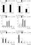
Figure 1. Altered USV production in old Tau.P301L mice.
A1- Rough USV spectrographic display with frequency and time scales in kHz and ms, respectively. A2 – As above but showing the tags (Tg, white dots) placed at slope changes in frequency on the rough USV and the analyzed USV parameters: duration of USV (Dur(USV)), frequency at tags (Freq(USV)), max and min Freq(USV) (RangeFreq), complexity (number of segments delimited by tags), and number of USV produced per min of recording (Nb(USV)). The total time of USV per min (totT(USV)) was obtained by summation of individual Dur(USV). B – Columns in histograms show Dur(USV) (expressed in ms) in Tau.P301L (black columns) and FVB/N (white columns) mice at age 4–5 months and 8–10 months (young and old mice, respectively). Note the significant reduction of Dur(USV) in old Tau.P301L mice. C – As in B but for Nb(USV) (expressed in USV per min). Note the drastic reduction of Nb(USV) in old Tau.P301L mice. D – As in B but totT(USV) (expressed in s per min of recording). Note the significant and drastic reduction of totT(USV) in old Tau.P301L mice. * indicates a significant inter-strain difference at a given class of age and $ a significant age-related difference for a given strain; ns, non significant inter-strain difference.
Figure 2
Figure 2. Altered USV pattern in old Tau.P301L mice.
A – Columns in histograms show Freq(USV) (expressed in kHz) used to produce USV in Tau.P301L (black columns) and FVB/N (white columns) mice at age 4–5 and 8–10 months. Note Freq(USV) was similar in old Tau.P301L and FVB/N mice. B – As in A, but for RangeFreq ( = max Freq(USV)−min Freq(USV), see Fig. 1A2). Note RangeFreq of old Tau.P301L mice was significantly reduced when compared to that of old FVB/N and that of young Tau.P301L mice. C - Columns in histograms show the distribution of totT(USV) (in %) vs. frequency (Freq), expressed in class of 10 kHz from 30 to 80 kHz in young Tau.P301L and FVB/N at age 4–5 months. Note that most USV used high 50–60 kHz frequency but some used low 30–40 kHz frequency in both genotypes. D – As in C, but for 8–10 months old mice. Arrows highlight that old Tau.P301L mice never used the low 30–40 kHz frequency component in their USV whereas old FVB/N mice still used both low and high frequencies. E - Columns in histograms show the occurrence (%) of USV of different complexity level as defined by the number of segments (see tags in Fig. 1A2) within the USV. Complexity ranged from low (1) to high (5). Note the similar distribution of complexity in Tau.P301L and FVB/N young mice. F – As in E but for old mice. Note the increased occurrence of USV of low complexity and the reduced occurrence of USV of higher complexity (>1) in old Tau.P301L mice compared to old FVB/N and young Tau.P301L mice. Complexity of old FVB/N mice did not change when compared to that of young FVB/N mice. * indicates a significant inter-strain difference at a given class of age and $ a significant age-related difference for a given strain; ns, non significant inter-strain difference.
Figure 3
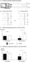
Figure 3. Reduced expiratory airflow in old Tau.P301L mice.
A - Schematic presentation of the double-chamber plethysmographic set-up allowing the simultaneous recordings of chest spirogram (CSp; in the body chamber) and airflow spirogram (ASp; in the head chamber) in conscious mice. B, C – Averaging of about 100 successive respiratory cycles during quiet period of breathing in young (B) and old (C) mice allowed the measurements of mean ASp and CSp, the calculation of the ASp/CSp ratio and the measurement of expiratory airflow during lung emptying period (gray areas). D - Columns in histograms show the ASp/CSp ratio in Tau.P301L (black columns) and FVB/N (white columns) young and old mice. Note 1) the ratio was similar in young Tau.P301L and FVB/N mice, and 2) the ratio was significantly reduced and increased in old Tau.P301L and FVB/N mice, respectively. E – As in D but for the expiratory airflow. Note the expiratory airflow was similar in young Tau.P301L and FVB/N mice, significantly halved in old Tau.P301L mice and unchanged in old FVB/N mice. * indicates a significant inter-strain difference at a given class of age and $ a significant age-related difference for a given strain; ns, non significant inter-strain difference.
Figure 4
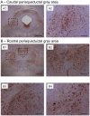
Figure 4. Tauopathy in the PAG of old Tau.P301L mice.
Immunohistochemistry with AT100 as tauopathy marker on midbrain coronal sections of old Tau.P301L mice reveals dramatic tauopathy in the whole PAG, affecting both its caudal (A) and rostral (B) parts. A2, B2 and B3 are enlargements of the dotted line boxes drawn in A1 and B1, and show high density of AT100+ neurons in the caudal, ventro-lateral PAG (A2), the rostral, dorso-median PAG (B2) and the rostral dorso-lateral PAG (B3) of the same old Tau.P301L mouse. B4 shows frequent AT100+ neurons in the rostral, dorso-lateral PAG of another old Tau.P301L mice. Calibration bars: 500 µm for A1, B1; 100 µm for A2, B2–B4.
Figure 5
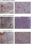
Figure 5. Tauopathy in the NRA and KF areas of old Tau.P301L mice.
Immunohistochemistry with AT100 as tauopathy marker on brainstem coronal sections of a given Tau.P301L mouse (as in Fig. 4). Right hand pictures are enlargements of the dotted line boxes drawn in left hand pictures. Sections show that AT100+ neurons are frequent in the NRA (A) and the KF (B) but lacking in the nucleus tractus solitarius and nucleus ambiguus (C). Calibration bars: 500 and 100 µm for left and right hand pictures, respectively. Labels in sections indicate the Kolliker-Fuse nucleus (KF), lateral parabrachial nucleus (LPB), nucleus ambiguus (nA), nucleus tractus solitarius (nTS), hypoglossal motor nucleus (n12), oral pontine reticular nucleus (PnO), principal trigeminal sensory nucleus (Pr5), pyramidal tract (Py), pyramidal decussation (Pyx), superior cerebella peduncle (scp), raphé dorsalis (RD), subcoeruelus nucleus (subC) and intra-medullary rootlet of hypoglossal nerve (12r).
Figure 6
Figure 6. Summary diagram of the organization of the PAG, KF and NRA network controlling vocalization.
Arrows indicate the demonstrated connections between structures controlling vocalization and numbers within arrows the related publications in the reference list. Mn, motor neurons; A6, locus coeruleus; RVLM, rostral ventrolateral medulla.

References

    1. Humbert IA, McLaren DG, Kosmatka K, Fitzgerald M, Johnson S, et al. Early deficits in cortical control of swallowing in Alzheimer's disease. J Alzheimers Dis. 2010;19:1185–1197. doi: 10.3233/JAD-2010-1316. - DOI - PMC - PubMed
    1. Suh MK, Kim H, Na DL. Dysphagia in patients with dementia: Alzheimer versus vascular. Alzheimer Dis Assoc Disord. 2009;23:178–184. doi: 10.1097/WAD.0b013e318192a539. - DOI - PubMed
    1. Attems J, König C, Huber M, Lintner F, Jellinger KA. Cause of death in demented and non-demented elderly inpatients; an autopsy study of 308 cases. J Alzheimers Dis. 2005;8:57–62. - PubMed
    1. Onen F, Onen H. [Obstructive sleep apnea and cognitive impairment in the elderly]. Psychol Neuropsychiatr Vieil. 2010;8:163–169. doi: 10.1684/pnv.2010.0219. - DOI - PubMed
    1. Cooke JR, Liu L, Natarajan L, He F, Marler M, et al. The effect of sleep-disordered breathing on stages of sleep in patients with Alzheimer's disease. Behav Sleep Med. 2006;4:219–27. doi: 10.1207/s15402010bsm0404_2. - DOI - PubMed

Publication types

MeSH terms