. 2011;6(10):e26125.
doi: 10.1371/journal.pone.0026125. Epub 2011 Oct 12.

Platelet adhesion and degranulation induce pro-survival and pro-angiogenic signalling in ovarian cancer cells

Affiliations

Platelet adhesion and degranulation induce pro-survival and pro-angiogenic signalling in ovarian cancer cells

Karl Egan et al. PLoS One. 2011.

Abstract

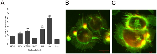
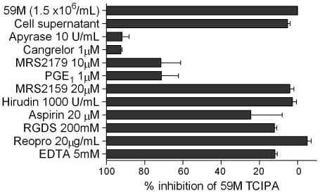
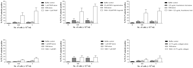
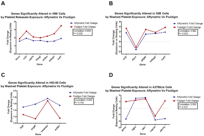
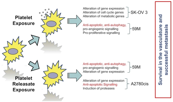
Thrombosis is common in ovarian cancer. However, the interaction of platelets with ovarian cancer cells has not been critically examined. To address this, we investigated platelet interactions in a range of ovarian cancer cell lines with different metastatic potentials [HIO-80, 59M, SK-OV-3, A2780, A2780cis]. Platelets adhered to ovarian cancer cells with the most significant adhesion to the 59M cell line. Ovarian cancer cells induced platelet activation [P-selectin expression] in a dose dependent manner, with the most significant activation seen in response to the 59M cell line. The platelet antagonists [cangrelor, MRS2179, and apyrase] inhibited 59M cell induced activation suggesting a P2Y12 and P2Y1 receptor mediated mechanism of platelet activation dependent on the release of ADP by 59M cells. A2780 and 59M cells potentiated PAR-1, PAR-4, and TxA2 receptor mediated platelet activation, but had no effect on ADP, epinephrine, or collagen induced activation. Analysis of gene expression changes in ovarian cancer cells following treatment with washed platelets or platelet releasate showed a subtle but valid upregulation of anti-apoptotic, anti-autophagy pro-angiogenic, pro-cell cycle and metabolic genes. Thus, ovarian cancer cells with different metastatic potential adhere and activate platelets differentially while both platelets and platelet releasate mediate pro-survival and pro-angiogenic signals in ovarian cancer cells.


Conflict of interest statement

Competing Interests: The authors have declared that no competing interests exist.

Figures

Figure 1
Figure 1. Platelet adhesion to a panel of ovarian cancer cell lines is heterogeneous under static conditions.
[A] Platelet adhesion to ovarian cancer cells was quantified based on the fluorescence detection of labelled platelets. Platelet adhesion to fibrinogen and BSA were used as ± controls [n = 8 + SEM, *  =  p<0.05 vs. BSA]. Fluorescence microscopy images of platelets adhering to A2780 [B] and 59M [C] cells under static conditions [representative of n = 3]. Ovarian cancer cells and platelets were stained for actin [green], platelets were stained specifically for CD42a [red/yellow].
Figure 2
Figure 2. Ovarian cancer cell lines induce platelet activation in a dose dependent manner [n = 3–6, ± SEM].
Platelet activation [P-selectin expression] induced by a range of ovarian cell lines over a large concentration range [0.1–1.5×10⁶/ml] was measured by flow cytometry, based on platelet P-selectin surface expression. The most significant platelet activation was seen in response to the metastatic ovarian cancer cell lines SK-OV-3 and 59M.
Figure 3
Figure 3. 59M tumour cell induced platelet activation (TCIPA) is inhibited by cangrelor, MRS2179, and apyrase.
This suggests a mechanism of platelet activation dependant on the platelet receptors P2Y12 and P2Y1, and ADP released by 59M cells into their supernatant. Other platelet antagonists such as hirudin, EDTA, abciximab, RGDS, and aspirin had no effect on 59M cell induced platelet activation.
Figure 4
Figure 4. 59M cells potentiate PAR-1, PAR-4, and TxA2 receptor mediated platelet activation.
Activation occurs in a dose dependent manner [n = 3, + SEM]. At low concentrations, 59M [0.5–1×10⁵/ml], cells significantly potentiated TRAP, PAR4 agonist, and Arachidonic acid induced platelet activation [P-selectin expression]. This suggests a synergistic relationship between PAR-1, PAR-4, and TxA2 receptor mediated platelet activation and 59M induced platelet activation. Similar results are seen for A2780 cells [1–5×10⁵ cells/ml, data not shown]
Figure 5
Figure 5. Fluidigm Dynamic Array Validation of significantly altered gene expression [fold change >1.5X; <1.5X; p<0.05].
This figure displays some examples of the correlation between the affymetrix array data and the fluidigm validation data. Correlation coefficients are displayed in the legend.
Figure 6
Figure 6. Summary analysis of gene expression changes due to platelets or platelet releasate.
Ovarian cancer cells exposed to platelets and platelet releasate exhibit a variety of gene expression changes summarised in this figure.
Figure 7
Figure 7. Volcano plot of TaqMan derived fold changes in EMT associated genes in cloaked ovarian cells.
This figure displays fold change difference in relation to p value for EMT genes assessed in the cell lines.

References

    1. Landen CN, Birrer MJ, Sood AK. Early events in the pathogenesis of epithelial ovarian cancer. J Clin Oncol. 2008;26:995–1005. - PubMed
    1. Holschneider CH, Berek JS. Ovarian cancer: epidemiology, biology, and prognostic factors. Semin Surg Oncol. 2000;19:3–10. - PubMed
    1. Naora H, Montell DJ. Ovarian cancer metastasis: integrating insights from disparate model organisms. Nat Rev Cancer. 2005;5:355–66. - PubMed
    1. Dvoretsky PM, Richards KA, Angel C, Rabinowitz L, Stoler MH, et al. Distribution of disease at autopsy in 100 women with ovarian cancer. Hum Pathol. 1988;19:57–63. - PubMed
    1. Braun S, Schindlbeck C, Hepp F, Janni W, Kentenich C, et al. Occult tumor cells in bone marrow of patients with locoregionally restricted ovarian cancer predict early distant metastatic relapse. J Clin Oncol. 2001;19:368–75. - PubMed

Publication types

MeSH terms