. 2011;6(10):e26180.
doi: 10.1371/journal.pone.0026180. Epub 2011 Oct 12.

Infection of XC cells by MLVs and Ebola virus is endosome-dependent but acidification-independent

Affiliations

Infection of XC cells by MLVs and Ebola virus is endosome-dependent but acidification-independent

Haruka Kamiyama et al. PLoS One. 2011.

Abstract

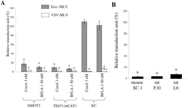
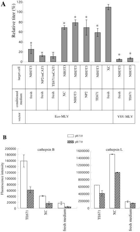
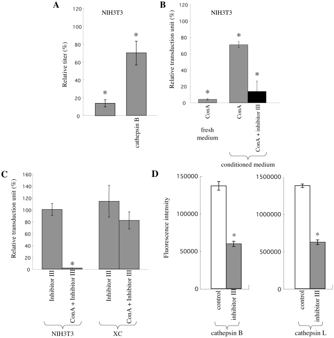
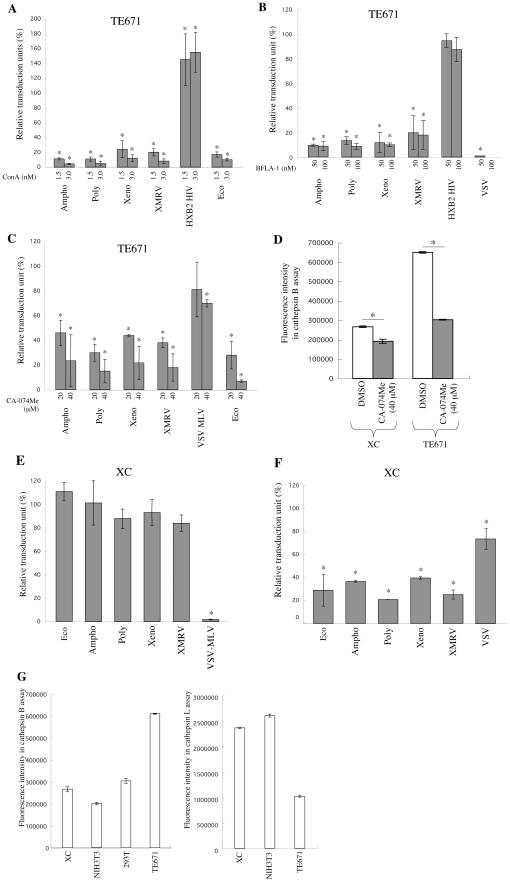
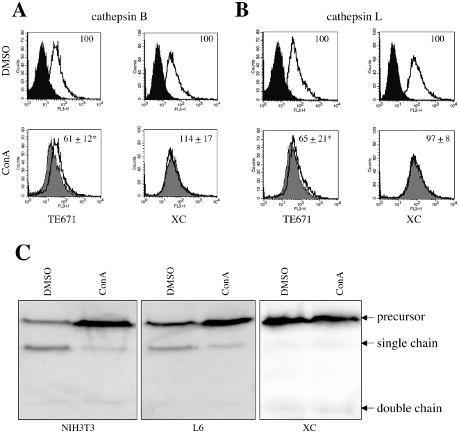
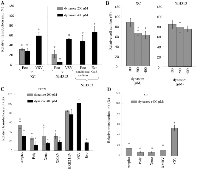
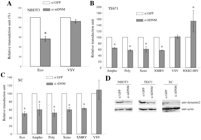
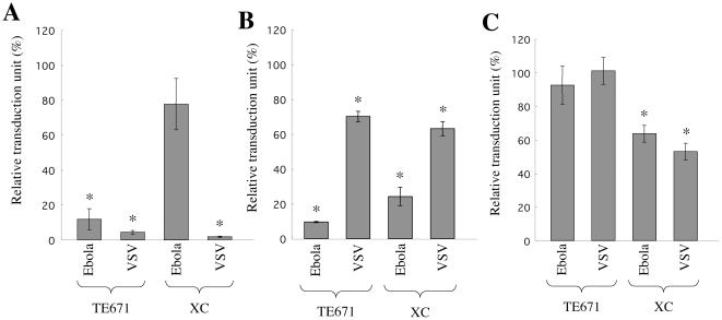
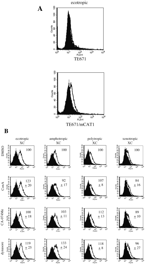
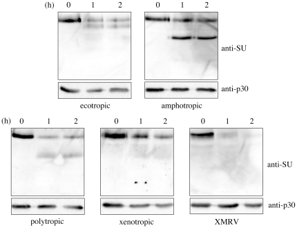
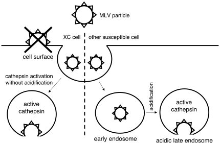
Inhibitors of endosome acidification or cathepsin proteases attenuated infections mediated by envelope proteins of xenotropic murine leukemia virus-related virus (XMRV) and Ebola virus, as well as ecotropic, amphotropic, polytropic, and xenotropic murine leukemia viruses (MLVs), indicating that infections by these viruses occur through acidic endosomes and require cathepsin proteases in the susceptible cells such as TE671 cells. However, as previously shown, the endosome acidification inhibitors did not inhibit these viral infections in XC cells. It is generally accepted that the ecotropic MLV infection in XC cells occurs at the plasma membrane. Because cathepsin proteases are activated by low pH in acidic endosomes, the acidification inhibitors may inhibit the viral infections by suppressing cathepsin protease activation. The acidification inhibitors attenuated the activities of cathepsin proteases B and L in TE671 cells, but not in XC cells. Processing of cathepsin protease L was suppressed by the acidification inhibitor in NIH3T3 cells, but again not in XC cells. These results indicate that cathepsin proteases are activated without endosome acidification in XC cells. Treatment with an endocytosis inhibitor or knockdown of dynamin 2 expression by siRNAs suppressed MLV infections in all examined cells including XC cells. Furthermore, endosomal cathepsin proteases were required for these viral infections in XC cells as other susceptible cells. These results suggest that infections of XC cells by the MLVs and Ebola virus occur through endosomes and pH-independent cathepsin activation induces pH-independent infection in XC cells.


Conflict of interest statement

Competing Interests: The authors have declared that no competing interests exist.

Figures

Figure 1
Figure 1. Eco-MLV infection in XC cells is independent of endosome acidification.
(A) Relative titers of the Eco- or VSV-MLV vector on ConA- or BFLA-1-treated NIH3T3, TE671/mCAT1, and XC cells were indicated. (B) Relative titers of the Eco-MLV vector on ConA-treated mouse SC-1, rat F10, and L6 cells were indicated. The transduction units obtained with DMSO-treated cells were set to 100%. These experiments were repeated in triplicate, and results are shown as the mean +/− SD. Asterisks indicate statistically significant differences compared to the value from the DMSO-treated cells.
Figure 2
Figure 2. Conditioned media induce pH-independent Eco-MLV infection.
(A) NIH3T3 cells were pretreated with ConA, and were inoculated with the Eco-MLV vector diluted with fresh or conditioned media. The transduction units obtained with DMSO-treated cells were set to 100%. These experiments were repeated in triplicate, and results are shown as the mean +/− SD. Asterisks indicate statistically significant differences compared to the value from the DMSO-treated cells. (B) Cathepsin B (left panel) and L (right panel) activities of TE671 and XC cell conditioned media were measured at pH 5.0 or 7.0. Fluorescence intensities were indicated. This experiment was repeated three times, and results are shown as means +/− SD.
Figure 3
Figure 3. Secreted cathepsin proteases induce pH-independent Eco-MLV infection.
(A) NIH3T3 cells were pretreated with ConA, and were inoculated with Eco-MLV vector diluted with fresh medium containing purified cathepsin B (100 ng/ml). (B) NIH3T3 cells were pretreated with ConA, and were inoculated with the Eco-MLV vector diluted with fresh or TE671 cell conditioned medium in the presence of cathepsin inhibitor III (200 µM). (C) NIH3T3 and XC cells were pretreated with ConA (3 nM) or cathepsin inhibitor III (200 µM). The treated cells were inoculated with Eco-MLV vector diluted with fresh medium in presence of cathepsin inhibitor III (200 µM). The transduction units obtained with DMSO-treated cells were set to 100%. These experiments were repeated in triplicate, and results are shown as the mean +/− SD. Asterisks indicate statistically significant differences compared to the value from the DMSO-treated cells. (D) Cathepsin B (left panel) and L (right panel) activities of TE671 cell conditioned medium in absence or presence of cathepsin inhibitor III were measured. Fluorescence intensities were indicated. This experiment was repeated three times, and results are shown as means +/− SD.
Figure 4
Figure 4. Ampho-, Poly-, Xeno-, and XMRV-MLV vector infections require endosome acidification and cathepsin proteases.
(A) TE671, TE671/mCAT1, or TE671/CD4 cells were pretreated with ConA for 5 h and were inoculated with the Eco-, Ampho-, Poly-, Xeno-, XMRV-MLV, or HXB2-HIV-1 vector. (B) TE671 or TE671/CD4 cells were pretreated with BFLA-1, and were inoculated with the Ampho-, Poly-, Xeno-, VSV-, XMRV-, VSV-MLV, or HXB2-HIV-1 vector. (C) TE671 or TE671/mCAT1 cells were pretreated with CA-074Me and were inoculated with the Eco-, Ampho-, Poly-, Xeno-, VSV-, or XMRV-MLV vector. The transduction units obtained with DMSO-treated cells were set to 100%. These experiments were repeated in triplicate, and results are shown as the mean +/− SD. Asterisks indicate statistically significant differences compared to the value from the DMSO-treated cells. (D) Cathepsin B activity of cells lysates from CA-074Me-treated cells was measured. Fluorescence intensities are shown. XC cells were treated with ConA (panel E) or CA-074Me (panel F), and were inoculated with the Eco-, Ampho-, Poly-, Xeno-, XMRV-, or VSV-MLV vector. These experiments were repeated in triplicate, and results are shown as the mean +/− SD. Asterisks indicate statistically significant differences compared to the value from the DMSO-treated cells. (G) Activities of cathepsins B (left panel) and L (right panel) were measured in cell lysates from XC, NIH3T3, 293T, and TE671 cells. Fluorescence intensities were indicated. These experiments were repeated three times, and results are shown as the mean +/− SD.
Figure 5
Figure 5. Treatment with endosome acidification inhibitors does not alter activities of cathepsins B and L in XC cells.
TE671 and XC cells were treated with ConA (3 nM) or DMSO. The treated cells were stained with the cathepsin B (panel A) or L (panel B) detection reagent. Closed areas indicate cells unstained with the reagents. Open areas indicate cells treated with DMSO and stained with the reagent. Grey areas indicate cells treated with ConA and stained with the reagent. Means of fluorescent intensity obtained with DMSO-treated cells were set to 100%. These experiments were repeated in triplicate, and results are shown as the mean +/− SD. Asterisks indicate statistically significant differences compared to the DMSO-treated cells. Mouse NIH3T3, rat L6, and XC cells were treated with DMSO or ConA (3 nM). Cell lysates from the treated cells were analyzed by Western immunoblotting using an anti-mouse cathepsin L ancbody (panel C).
Figure 6
Figure 6. MLV infections are inhibited by dynasore.
(A) XC and NIH3T3 cells were pretreated with dynasore and were inoculated with the Eco-MLV vector diluted with fresh, TE671 cell conditioned, or cathepsin B (100 ng/ml)-containing medium. (B) XC and NIH3T3 cells were treated with dynasore, washed, and cultured for 48 h. Cells resistant to trypan blue staining were quantified. (C) TE671, TE671/mCAT1, or TE671/CD4 cells were pretreated with dynasore and were inoculated with the Eco-, Ampho-, Poly-, Xeno-, XMRV-, VSV-MLV, or HXB2-HIV-1 vector. (D) XC cells were pretreated with dynasore and were inoculated with the Ampho-, Poly-, Xeno-, XMRV-, or VSV-MLV vector. The transduction units obtained with DMSO-treated cells were set to 100%. These experiments were repeated in triplicate, and results are shown as the mean +/− SD. Asterisks indicate statistically significant differences compared to the value from the DMSO-treated cells.
Figure 7
Figure 7. MLV infections are inhibited by siRNA-mediated knockdown of dynamin 2 expression.
(A) NIH3T3 cells were transfected with an siRNA against mouse dynamin 2 mRNA, and were inoculated with the Eco-MLV vector. (B) TE671 or TE671/CD4 cells were transfected with an siRNA against human dynamin 2 mRNA, and were inoculated with the Ampho-, Poly-, Xeno-, XMRV-, VSV-MLV, or HXB2-HIV-1 vector. (C) XC cells were transfected with an siRNA against rat dynamin 2 mRNA, and were inoculated with the Eco-, Ampho-, Poly-, Xeno-, XMRV- or VSV-MLV vector. The transduction units obtained with DMSO-treated cells were set to 100%. These experiments were repeated in triplicate, and results are shown as the mean +/− SD. Asterisks indicate statistically significant differences compared to the value from the DMSO-treated cells. (D) Dynamin 2 protein expression in the siRNA-transfected cells was analyzed by Western immunoblotting using an anti-dynamin 2 antibody. Actin expression was analyzed as control.
Figure 8
Figure 8. Ebola virus infection in XC cells is pH-independent.
TE671 and XC cells were pretreated with ConA (panel A), CA-074Me (panel B), or dynasore (panel C), and were inoculated with the Ebola virus GP- or VSV-G-pseudotyped MLV vector. The transduction units obtained with DMSO-treated cells were set to 100%. These experiments were repeated in triplicate, and results are shown as the mean +/− SD. Asterisks indicate statistically significant differences compared to the value from the DMSO-treated cells.
Figure 9
Figure 9. Binding of the MLV vectors to target cells is not affected by ConA, CA-074Me, or dynasore treatments.
(A) Binding of the Eco-MLV vector particles to TE671 (upper panel) and TE671/mCAT1 (lower panel) was analyzed. (B) XC cells were pretreated with DMSO, ConA, CA-074Me, or dynasore, and were incubated with the Eco-, Ampho-, Poly-, or Xeno-MLV vector. The fluorescence intensities obtained with DMSO-treated cells were set to 100%. These experiments were repeated in triplicate, and values are shown as the mean +/− SD.
Figure 10
Figure 10. Digestion of MLV Env proteins by cathepsin B.
The MLV vector particles were treated with cathepsin B (100 ng/ml). The products were analyzed by Western immunoblotting using the anti-MLV SU antiserum.
Figure 11
Figure 11. Entry pathways of MLV infection in XC cells and in other susceptible cells.

References

    1. Albritton LM, Tseng L, Scadden D, Cunningham JM. A putative murine ecotropic retrovirus receptor gene encodes a multiple membrane-spanning protein and confers susceptibility to virus infection. Cell. 1989;57:659–666. - PubMed
    1. Kavanaugh MP, Miller DG, Zhang W, Law W, Kozak SL, et al. Cell-surface receptors for gibbon ape leukemia virus and amphotropic murine retrovirus are inducible sodium-dependent phosphate symporters. Proc Natl Acad Sci U S A. 1994;91:7071–7075. - PMC - PubMed
    1. Miller DG, Edwards RH, Miller AD. Cloning of the cellular receptor for amphotropic murine retroviruses reveals homology to that for gibbon ape leukemia virus. Proc Natl Acad Sci U S A. 1994;91:78–82. - PMC - PubMed
    1. van Zeijl M, Johann SV, Closs E, Cunningham J, Eddy R, et al. A human amphotropic retrovirus receptor is a second member of the gibbon ape leukemia virus receptor family. Proc Natl Acad Sci U S A. 1994;91:1168–1172. - PMC - PubMed
    1. Battini JL, Rasko JE, Miller AD. A human cell-surface receptor for xenotropic and polytropic murine leukemia viruses: possible role in G protein-coupled signal transduction. Proc Natl Acad Sci U S A. 1999;96:1385–1390. - PMC - PubMed

Publication types

MeSH terms