. 2011;6(10):e26187.
doi: 10.1371/journal.pone.0026187. Epub 2011 Oct 13.

Synergistic NGF/B27 gradients position synapses heterogeneously in 3D micropatterned neural cultures

Affiliations

Synergistic NGF/B27 gradients position synapses heterogeneously in 3D micropatterned neural cultures

Anja Kunze et al. PLoS One. 2011.

Abstract

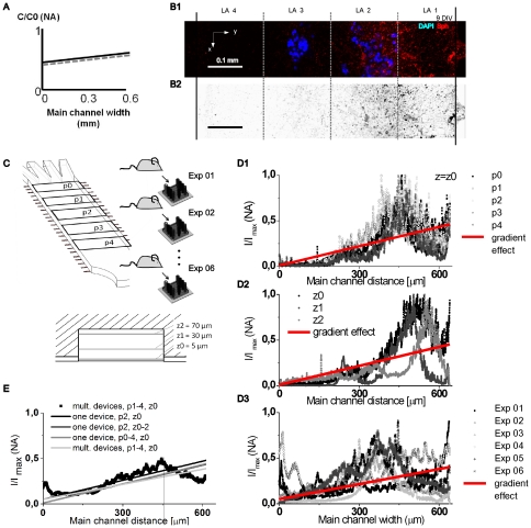
Native functional brain circuits show different numbers of synapses (synaptic densities) in the cerebral cortex. Until now, different synaptic densities could not be studied in vitro using current cell culture methods for primary neurons. Herein, we present a novel microfluidic based cell culture method that combines 3D micropatterning of hydrogel layers with linear chemical gradient formation. Micropatterned hydrogels were used to encapsulate dissociated cortical neurons in laminar cell layers and neurotrophic factors NGF and B27 were added to influence the formation of synapses. Neurotrophic gradients allowed for the positioning of distinguishable synaptic densities throughout a 3D micropatterned neural culture. NGF and B27 gradients were maintained in the microfluidic device for over two weeks without perfusion pumps by utilizing a refilling procedure. Spatial distribution of synapses was examined with a pre-synaptic marker to determine synaptic densities. From our experiments, we observed that (1) cortical neurons responded only to synergistic NGF/B27 gradients, (2) synaptic density increased proportionally to synergistic NGF/B27 gradients; (3) homogeneous distribution of B27 disturbed cortical neurons in sensing NGF gradients and (4) the cell layer position significantly impacted spatial distribution of synapses.


Conflict of interest statement

Competing Interests: The authors have declared that no competing interests exist.

Figures

Figure 1
Figure 1. Engineering spatial distribution of synapses in microfabricated 3D neural cell layers.
(A) Illustration of native synaptic appearance in the cerebral cortex. Scale adapted to rats. L1 … L6: cortical cell layer notation. (B) Native synaptic density differs within cortical cell layers. Synaptic density was extracted from based on image treatment, described in Supporting information S6. We hypothesize that synergistic NGF/B27 gradients influence axon guidance and spatial distribution of synapses. (C) Schematic view of combining micropatterning and gradient generation in a polydimethylsiloxane (PDMS) microfluidic device. (D) Engineered cortical cell layers and spatial distribution of synapses after B27/NGF gradient exposure. Synergistic gradient guides neurites and increases synapse assembly towards higher concentration. LA 1 … LA 4: hydrogel layer notation in vitro.
Figure 2
Figure 2. Establishing stable long-term gradients through refilling.
(A) Illustration of reservoir refilling procedure. Step 1, empty polydimethylsiloxane (PDMS) reservoirs are selectively filled with medium. Red color indicates enriched NGF/B27 condition. Green color represents pure medium. Stable linear gradients establishes through junction channels and micropatterned hydrogel layers in the main channel, because of perfusion flow. Step 2, after 2 h perfusion flow stops. The long perfusion channels maintain the gradient in the main channel. Dimensions are in mm. Every other day, refilling was repeated. (B) Experimental gradient formation and computational adaption of reduced diffusion in the hydrogel layers in the main channel. (C) Computational stable NGF gradient formation over cell culture period. ∇C stabilizes after 2 h, whereas Cavg reaches stable point after 2 d, but before dissociated neurons response.
Figure 3
Figure 3. Neurite outgrowth and guidance towards synergistic B27 and NGF gradient.
(A, row) Schematic view of single versus synergistic NGF and B27 gradients, which stimulate micropatterned cell cultures in the main channel. (B, row) Differential interference contrast (DIC) images of micropatterned neural cell culture (E19) after 9 days in vitro (DIV), bar  = 0.1 mm. (C, row) Inverted DIC images with traced neurites, bar  = 0.1 mm. (D, row) Neurite lengths grown in left versus right hydrogel layers. (E, row) Neurite traces from 2 and 9 DIV, summarized in polar plots. Lengths of radii are in µm and angles are in degree. Only synergistic NGF/B27 orient neurite outgrowth towards higher concentrations.
Figure 4
Figure 4. Using synergistic NGF/B27 gradients polarizes spatial synapse distribution towards higher concentrations.
(A) Schematic view of synergistic NGF/B27 gradients in the main channel. (B1) False color images shows micropatterned cell layers through nuclei staining (DAPI, blue) and polarized pre-synaptic units (Synaptophysin, red). (B2) Inverted red channel highlight synapse distribution. (C) Evaluation parameters. (D, column) Surface plot of spatial synapse distribution and linear regression fit of data. D1: different lateral positions, D2: different vertical positions and D3: different experimental batches. (E) Averaged spatial synapse distribution correlates with linear fit of data (gradient effect) and is independent of evaluation parameters.
Figure 5
Figure 5. Different combinations of synergistic NGF/B27 gradients impact spatial synapse distribution.
(A and C) Gradient input in the main channel. (A) Stable NGF gradient was combined with a homogenous B27 distribution or a B27 gradient. (B and D) Linear regression fits from synapse distribution. (B) Homogenous B27 distribution disturbs NGF gradient sensing. Cortical neurons express polarized synapse distribution only when synergistic NGF/B27 gradients interplay. (C) Homogenous B27 distribution was combined with increased ∇C/Cavg NGF gradients. (D) Disturbed synapse distribution was recovered through high relative NGF gradients.
Figure 6
Figure 6. Cell layer position influences spatial synapse distribution.
(A1 and B1) Schematic view of shifted cell layer position and NGF/B27 (12B27pNGF-53) gradient exposure. (A2 and B2) False color image shows cell pattern dependent synapse distribution in the micropatterned cell culture after 9 DIV. Pre-synaptic units: synaptophysin (red), cell nucleus: DAPI (blue), scale bar  = 100 µm. (A3 and B3) Surface plots of synapse distribution with linear fit of data demonstrate synaptic gradient response, z  = 5 µm.

References

    1. Lefort S, Tomm C, Floyd Sarria JC, Petersen CCH. The Excitatory Neuronal Network of the C2 Barrel Column in Mouse Primary Somatosensory Cortex. Neuron. 2009;61:301–316. - PubMed
    1. Somogyi P, Freund TF, Cowey A. The axo-axonic interneuron in the cerebral cortex of the rat, cat and monkey. Neuroscience. 1982;7:2577–2607. - PubMed
    1. Micheva KD, Busse B, Weiler NC, O'Rourke N, Smith SJ. Single-Synapse Analysis of a Diverse Synapse Population: Proteomic Imaging Methods and Markers. Neuron. 2010;68:639–653. - PMC - PubMed
    1. Shen K, Cowan CW. Guidance Molecules in Synapse Formation and Plasticity. Cold Spring Harbor Perspectives in Biology. 2010;2 - PMC - PubMed
    1. Sofroniew M, Howe C, Mobley W. Nerve growth factor signaling, neuroprotection, and neural repair. Annu Rev Neurosci. 2001;24:1217–1281. - PubMed

Publication types

Substances