. 2011;6(10):e26218.
doi: 10.1371/journal.pone.0026218. Epub 2011 Oct 14.

Identification of intracellular and plasma membrane calcium channel homologues in pathogenic parasites

Affiliations

Identification of intracellular and plasma membrane calcium channel homologues in pathogenic parasites

David L Prole et al. PLoS One. 2011.

Abstract

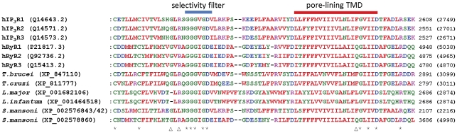
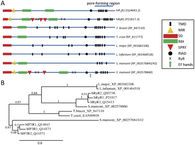
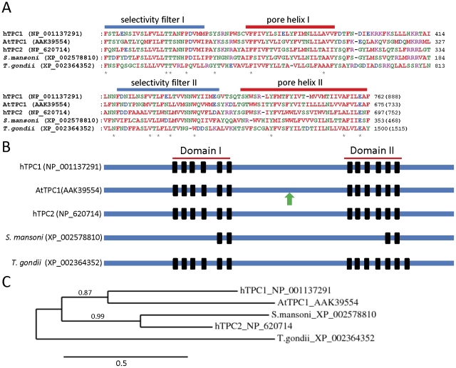
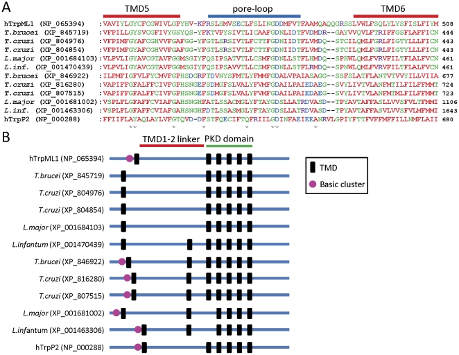
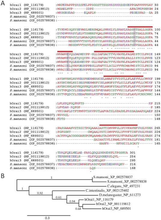
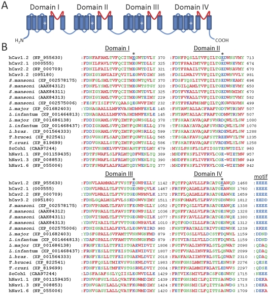
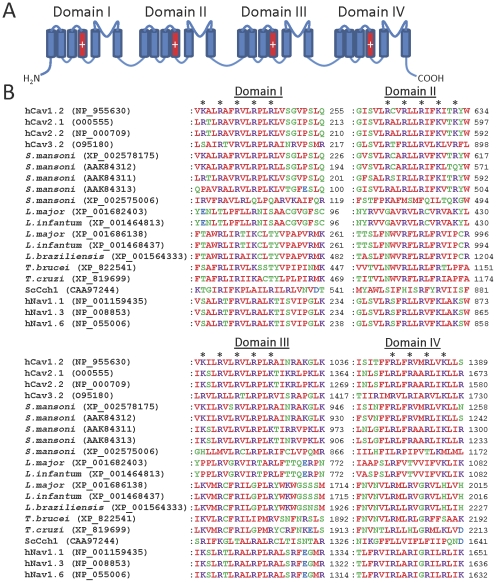
Ca(2+) channels regulate many crucial processes within cells and their abnormal activity can be damaging to cell survival, suggesting that they might represent attractive therapeutic targets in pathogenic organisms. Parasitic diseases such as malaria, leishmaniasis, trypanosomiasis and schistosomiasis are responsible for millions of deaths each year worldwide. The genomes of many pathogenic parasites have recently been sequenced, opening the way for rational design of targeted therapies. We analyzed genomes of pathogenic protozoan parasites as well as the genome of Schistosoma mansoni, and show the existence within them of genes encoding homologues of mammalian intracellular Ca(2+) release channels: inositol 1,4,5-trisphosphate receptors (IP(3)Rs), ryanodine receptors (RyRs), two-pore Ca(2+) channels (TPCs) and intracellular transient receptor potential (Trp) channels. The genomes of Trypanosoma, Leishmania and S. mansoni parasites encode IP(3)R/RyR and Trp channel homologues, and that of S. mansoni additionally encodes a TPC homologue. In contrast, apicomplexan parasites lack genes encoding IP(3)R/RyR homologues and possess only genes encoding TPC and Trp channel homologues (Toxoplasma gondii) or Trp channel homologues alone. The genomes of parasites also encode homologues of mammalian Ca(2+) influx channels, including voltage-gated Ca(2+) channels and plasma membrane Trp channels. The genome of S. mansoni also encodes Orai Ca(2+) channel and STIM Ca(2+) sensor homologues, suggesting that store-operated Ca(2+) entry may occur in this parasite. Many anti-parasitic agents alter parasite Ca(2+) homeostasis and some are known modulators of mammalian Ca(2+) channels, suggesting that parasite Ca(2+) channel homologues might be the targets of some current anti-parasitic drugs. Differences between human and parasite Ca(2+) channels suggest that pathogen-specific targeting of these channels may be an attractive therapeutic prospect.


Conflict of interest statement

Competing Interests: The authors have declared that no competing interests exist.

Figures

Figure 1
Figure 1. Alignment of parasite putative IP3Rs/RyRs with the pore regions of mammalian IP3Rs/RyRs.
Alignments of the pore region of human IP3Rs and RyRs with putative homologues from T. brucei, T. cruzi, L. infantum, and S. mansoni. ClustalW2 physiochemical residue colours are shown, and protein accession numbers are shown in parentheses after the species name. Numbers in parentheses to the right of the alignments indicate the total number of residues in the protein. Asterisks below the alignment indicate absolute residue conservation in all homologues. Triangles below the alignment indicate the position of residues discussed in the text (G4891, R4893 and Q4934 of hRyR1). The blue bar above the alignment indicates the selectivity filter region, while the red bar indicates the pore-forming transmembrane domain (TMD). The S. mansoni protein labelled XP_002576843/42 is formed by concatenated XP_002576843 and XP_002576842 predicted proteins (whose genomic loci are adjacent). The entire concatamer was confirmed as a single intact protein by its identity with the predicted protein: 29191.m000804.twinscan2 (Wellcome Trust Sanger Institute, UK).
Figure 2
Figure 2. Comparison of full-length sequences of human and parasite IP3R/RyR homologues.
(A) Schematic showing location of putative transmembrane domains (TMDs) and conserved domains identified using the Conserved Domains Database (NCBI), including: MIR (mannosyltransferase, IP3R and RyR) domains (pfam02815), RIH (RyR and IP3R homology) domains (pfam01365), RIH-associated domains (RIAD) (pfam08454), EF hands, suppressor-domain-like domains (SD) (pfam08709), RyR (ryanodine receptor) domains (pfam02026), and SPRY (SPla and the ryanodine receptor) domains (pfam00622). Putative pore-forming regions (pfam00520) are also indicated. (B) Phylogram showing relationships between full-length sequences of human and parasite IP3R/RyR homologues (see Methods: based on 226 high confidence positions from a multiple sequence alignment; gamma shape parameter 1.755; proportion of invariant sites 0). Branch length scale bar and branch support values are shown.
Figure 3
Figure 3. Comparison of human, plant and parasite TPC homologues.
(A) Alignments of pore domains I (upper panel) and II (lower panel) of human, plant (AtTPC1 is Arabidopsis thaliana TPC1) and parasite TPC homologues. Asterisks below the alignment indicate absolute residue conservation in all homologues. (B) Schematic showing the location of transmembrane domains (black bars) within the full-length sequences of human, plant and parasite TPC homologues. The green arrow signifies an EF-hand repeat in AtTPC1. (C) Phylogram showing the relationship between full-length sequences of human, plant and parasite TPC homologues (see Methods: based on 220 high confidence positions from a multiple sequence alignment; gamma shape parameter 4.195; proportion of invariant sites 0.021). Branch length scale bar and branch support values are shown.
Figure 4
Figure 4. Trp channel homologues in kinetoplastid parasites.
(A) Alignment of the pore regions of human TrpML and TrpP2 channel subunits with putative homologues from kinetoplastid parasites. Sequences of the putative pore loops as well as part of the TMD5 and TMD6 regions are shown. Numbers in parentheses to the right of the alignments indicate the total number of residues in the protein. Asterisks below the alignment indicate residues that are absolutely conserved between parasite proteins and either hTrpML1 or hTrpP2. L. infantum is shown abbreviated to L. inf. (B) Schematic showing the location of predicted TMDs (black bars) within the full-length sequences of human TrpML1 and TrpP2 channel subunits, as well as parasite homologues. The conserved PKD polycystin domain (pfam08016; identified using the Conserved Domains Database, NCBI) is indicated, as well as the long TMD1-TMD2 linker characteristic of mammalian TrpML and TrpP2 channel subunits. The presence of an N-terminal cluster of basic residues preceding TMD1 is indicated by a purple circle.
Figure 5
Figure 5. Comparison of human and S. mansoni Orai homologues.
(A) Alignments of human Orai subunits with predicted homologues from S. mansoni are shown (XP_002578838 is a shorter alternatively spliced variant of XP_002578837). Residues within the putative pore that are crucial for hOrai1 function (R91 and E106) are highlighted by grey bars. Predicted transmembrane regions of hOrai1 are indicated by red bars above the alignment, and asterisks below the alignment denote sequence identity amongst all isoforms. (B) Phylogram showing the relationship between human and S. mansoni Orai homologues as well as those of the model invertebrates C. elegans, D. melanogaster and C. intestinalis (see Methods: based on 141 high confidence positions from a multiple sequence alignment; gamma shape parameter 1.541; proportion of invariant sites 0.156). Branch length scale bar and branch support values are shown.
Figure 6
Figure 6. Parasite Cav channel homologues show similarity to human Cav channels in the pore region.
(A) Schematic showing the four-domain structure of human Cav channels, with the pore loop of each domain shown in red. Cylinders indicate TMDs, and plus signs indicate the charged voltage sensor regions. (B) Multiple sequence alignments of the pore domains of human Cav channels with parasite Cav channel homologues. Only those parasite homologues containing four putative domains are shown. Sequences of human Cav1.2 (L-type), Cav2.1 (P/Q-type), Cav2.2 (N-type) and Cav3.2 (T-type) channels, as well as the S. cerevisiae Cch1 Ca2+ channel (ScCch1), are shown. L. braz denotes L. braziliensis. Selected human Nav isoforms are included, to allow comparison with the related Cav channels. The locations of acidic residues (underlined) forming an acidic ring motif in human Cav1.2 channels are indicated by asterisks. The overall motif formed by all four domains at this locus is indicated to the right of the Domain IV alignment for each channel homologue.
Figure 7
Figure 7. Parasite Cav channel homologues show similarity to human Cav channels in the voltage sensor region.
(A) Schematic showing the four-domain structure of human Cav channels, with the voltage sensors of each domain shown in red. (B) Multiple sequence alignments of the voltage sensor domains of human Cav channels with parasite Cav channel homologues. Only those parasite homologues containing four putative domains are shown. Asterisks indicate the position of basic residues which form the voltage sensor in each domain of human Cav1.2 channels.

References

    1. Philosoph H, Zilberstein D. Regulation of intracellular calcium in promastigotes of the human protozoan parasite Leishmania donovani. J Biol Chem. 1989;264:10420–4. - PubMed
    1. Moreno SN, Docampo R. Calcium regulation in protozoan parasites. Curr Opin Microbiol. 2003;6:359–64. - PubMed
    1. Nagamune K, Moreno SN, Chini EN, Sibley LD. Calcium regulation and signaling in apicomplexan parasites. Subcell Biochem. 2008;47:70–81. - PubMed
    1. Billker O, Lourido S, Sibley LD. Calcium-dependent signaling and kinases in apicomplexan parasites. Cell Host Microbe. 2009;5:612–22. - PMC - PubMed
    1. Marks AR. Intracellular calcium-release channels: regulators of cell life and death. Am J Physiol. 1997;272:H597–605. - PubMed

Publication types

MeSH terms

Substances