. 2012 Jan;279(1):100-12.
doi: 10.1111/j.1742-4658.2011.08402.x. Epub 2011 Nov 11.

Biosynthesis of UDP-glucuronic acid and UDP-galacturonic acid in Bacillus cereus subsp. cytotoxis NVH 391-98

Affiliations

Biosynthesis of UDP-glucuronic acid and UDP-galacturonic acid in Bacillus cereus subsp. cytotoxis NVH 391-98

Bryan Broach et al. FEBS J. 2012 Jan.

Abstract

The food borne pathogen Bacillus cereus produces uronic acid-containing glycans that are secreted in a shielding biofilm environment, and certain alkaliphilic Bacillus deposit uronate-glycan polymers in the cell wall when adapting to alkaline environments. The source of these acidic sugars is unknown and, in the present study, we describe the functional identification of an operon in Bacillus cerues subsp. cytotoxis NVH 391-98 that comprises genes involved in the synthesis of UDP-uronic acids in Bacillus spp. Within the operon, a UDP-glucose 6-dehydrogenase converts UDP-glucose in the presence of NAD(+) to UDP-glucuronic acid and NADH, and a UDP-GlcA 4-epimerase (UGlcAE) converts UDP-glucuronic acid to UDP-galacturonic acid. Interestingly, in vitro, both enzymes can utilize the TDP-sugar forms as well, albeit at lower catalytic efficiency. Unlike most of the very few bacterial 4-epimerases that have been characterized, which are promiscuous, the B. cereus UGlcAE enzyme is very specific and cannot use UDP-glucose, UDP-N-acetylglucosamine, UDP-N-acetylglucosaminuronic acid or UDP-xylose as substrates. Size exclusion chromatography suggests that UGlcAE is active as a monomer, unlike the dimeric form of plant enzymes; the Bacillus UDP-glucose 6-dehydrogenase is also found as a monomer. Phylogenic analysis further suggests that the Bacillus UGlcAE may have evolved separately from other bacterial and plant epimerases. Our results provide insight into the formation and function of uronic acid-containing glycans in the lifecycle of B. cereus and related species containing homologous operons, as well as a basis for determining the importance of these acidic glycans. We also discuss the ability to target UGlcAE as a drug candidate.


Figures

FIGURE 1
FIGURE 1. The biosyntheses of acidic sugar nucleotides in Bacillus.
A. In Bacillus, based on the present report, UDP-Glc pyrophosphorylase (UDP-Glc PPase) converts Glc-1-P and UTP to UDP-Glc. In the presence of NAD+, UDP-Glc is interconverted by UDP-glucose dehydrogenase (UGlcDH) into UDP-GlcA and NADH. UDP-GlcA can then be interconverted by the 4-epimerase, UGlcAE, into UDP-GalA. B. Organization of the three genes within the UGalA operon and the flanking regions across selective members of Bacillus spp. The locus number for each gene in the operon across the different species shown is indicated. Based on this report, Bcer98_2076 functions as a UGlcDH and Bcer98_2077 as a UGlcAE. The operon also comprises a putative glycosyltransferase (Bcer98_2078 in B. cereus) which may be involved in the synthesis of GlcA or GalA-containing glycans. Note: except for Geobacillus sp. G11MC16, in the 5′ region flanking the UGalA operon there is a single gene in the opposite direction of the operon encoding a putative UDP-Glc PPase (Bcer98_2075 in B. cereus, putative ytdA in B. subtilis).
FIGURE 2
FIGURE 2. Expression and characterization of recombinant of BcUGlcDH
A. SDS-PAGE of total soluble protein isolated from E. coli cells expressing UGlcDH, (lane 2), negative vector control (lane 3), and of column-purified UGlcDH or control (lane 4 and 5, respectively). An arrow points to the purified protein. B. High performance anion-exchange chromatography of the products formed by UGlcDH. Purified recombinant UGlcDH was reacted with UDP-Glc for 30 min in the presence (Panel 4) or absence (Panel 6) of NAD+. The corresponding column-purified protein isolated from cells expressing the control vector was reacted with UDP-Glc and NAD+ for 30 min (Panel 5) as a control. The reaction products were separated on a Q15 anion-exchange column. The distinct UDP-sugar peak marked by an asterisk (Panel 2 and 4 with retention time of 18.8 min) was collected and characterized by mass spectrometry and 1H-NMR spectroscopy. The activity of total soluble protein (denoted S20) isolated from cells expressing recombinant BcUGlcDH (Panel 2) or vector control (Panel 3) is also shown. C. Analysis of BcUGlcDH enzymatic product by mass spectrometry operated in the negative ion mode. The HPLC peak (Panel 2B or 4B, marked by *) was collected and directly infused to ESI-MS. The spectrum of the full MS molecular ions (Panel 1, m/z of 579.05 for deprotonated [M-H]) and of the derived CID-fragments (Panel 2, m/z of 323.06 and 403.05) are shown. D. The peak in Panel 4B marked by ** has major UV absorbance peaks at 259 and 340 nm, the characteristic absorbance signature for NADH.
FIGURE 3
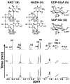
FIGURE 3. Analysis of BcUGlcDH enzymatic reaction in time-resolved 1H-NMR
Selected regions of the 600 mHz 1H-NMR spectrum show the interconversion of UDP-α-D-Glc and NAD+ to UDP-α-D-GlcA and NADH at 5, 15, and 45 min after the reaction was initiated. Diagnostic peaks of NAD+ and NADH are labeled N and H, respectively, while UDP-Glc peaks are labeled G and UDP-GlcA peaks are labeled A. Peaks are arbitrarily numbered to denote corresponding protons numbered as indicated in the molecular structures.
FIGURE 4
FIGURE 4. BcUGlcDH can convert TDP-Glc to TDP-GlcA as determined by MALDI and NMR
A. MALDI-MS analysis - Negative-ion MALDI mass spectra were recorded using a Microflex LT mass spectrometer (Bruker Daltonik, Bremen, Germany). 1 μl aqueous samples of 1 mM TDP-GlcA (produced from the dehydrogenase reaction with TDP-Glc/NAD+ and collected by HPLC) were mixed with an equalvolume of matrix solution (1 μg/μl 2,5-dihydroxybenzoic acid in 50% methanol) and driedon the plate. Spectra from 500 laser(N2, 337nm) shots were summed to generate a mass spectrum. The ion mass, calculated (abbr. Cal.) or observed (abbr. Obs.), are listed below the spectrum. B. 1H-NMR spectroscopy at 600-MHz of HPLC collected peak after reaction of BcUGlcDH with TDP-Glc and NAD+. The assignments of each proton are indicated on the spectrum: peaks from protons on the thymidine (T) ring are indicated by H, the ribose (R) protons are indicated by H′, and the GlcA (G) protons protons are indicated by H″. The peaks marked by * are resonances from impurities.
FIGURE 5
FIGURE 5. Expression and characterization of recombinant BcUGlcAE
A. SDS-PAGE of total soluble protein isolated from E. coli cells expressing BcUGlcAE (lane 1), control vector (lane 2), and of column-purified BcUGlcAE or control (lane 3 and 4, respectively). B. HPLC analysis of the products formed by BcUGlcAE reacting with UDP-GlcA. Total soluble proteins (denoted S20) or purified recombinant UGlcAE was reacted with UDP-GlcA for 30 min in the presence (Panel 2, 3), or absence (Panel 4) of NAD+. The crude or purified protein isolated from cells expressing control vector was incubated with UDP-GlcA for 60 min (Panel 5,6) as a control. The distinct UDP-sugar peak marked by the arrow (Panel 2, 3, and 4 with the same retention time) was collected and analyzed by 1H-NMR spectroscopy. C. HPLC analysis of BcUGlcAE that was reacted with TDP-GlcA to yield a product (marked by an arrow in panels 2, 3, and 4) that was collected and analyzed by NMR and MALDI-TOF.
FIGURE 6
FIGURE 6. 1H-NMR spectroscopic and MALDI-TOF analyses of BcUGlcAE reaction products indicates formation of TDP-α-D-galacturonic acid
A. The HPLC peaks (Fig. 5C, Panel 2, 3, or 4) corresponding to the product formed by BcUGlcAE was collected and analyzed by MALDI-TOF. B. 1H-NMR spectroscopy at 600-MHz of HPLC peak from Fig. 5C panel 3. The assignments of each proton are indicated on the spectrum: peaks from proto ns on the thymidine (T) ring are indicated by H, the ribose (R) protons are indicated by H′, and the GalA (G) protons protons are indicated by H″. The peaks marked by * are resonances from impurities.

References

    1. Leoff C, Saile E, Rauvolfova J, Quinn CP, Hoffmaster AR, Zhong W, Mehta AS, Boons GJ, Carlson RW, Kannenberg EL. Secondary cell wall polysaccharides of Bacillus anthracis are antigens that contain specific epitopes which cross-react with three pathogenic Bacillus cereus strains that caused severe disease, and other epitopes common to all the Bacillus cereus strains tested. Glycobiology. 2009;19:665–73. - PMC - PubMed
    1. Raetz CR, Whitfield C. Lipopolysaccharide endotoxins. Annu Rev Biochem. 2002;71:635–700. - PMC - PubMed
    1. Muszynski A, Laus M, Kijne JW, Carlson RW. Structures of the lipopolysaccharides from Rhizobium leguminosarum RBL5523 and its UDP-glucose dehydrogenase mutant (exo5) Glycobiology. 2011;21:55–68. - PMC - PubMed
    1. Quelas JI, Mongiardini EJ, Casabuono A, Lopez-Garcia SL, Althabegoiti MJ, Covelli JM, Perez-Gimenez J, Couto A, Lodeiro AR. Lack of galactose or galacturonic acid in Bradyrhizobium japonicum USDA 110 exopolysaccharide leads to different symbiotic responses in soybean. Molecular plant-microbe interactions: MPMI. 2010;23:1592–604. - PubMed
    1. Frirdich E, Whitfield C. Characterization of Gla(KP), a UDP-galacturonic acid C4-epimerase from Klebsiella pneumoniae with extended substrate specificity. J Bacteriol. 2005;187:4104–15. - PMC - PubMed

Publication types

MeSH terms

Substances

Associated data

LinkOut - more resources