. 2011 Nov 16;133(45):18328-42.
doi: 10.1021/ja207163r. Epub 2011 Oct 24.

Phosphorescent sensor for biological mobile zinc

Affiliations

Phosphorescent sensor for biological mobile zinc

Youngmin You et al. J Am Chem Soc. .

Abstract

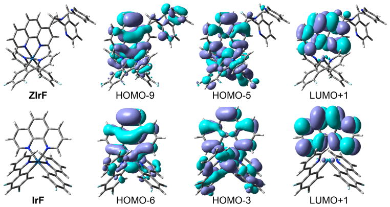
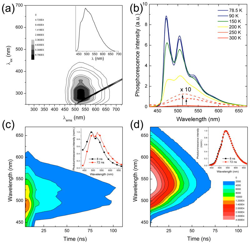
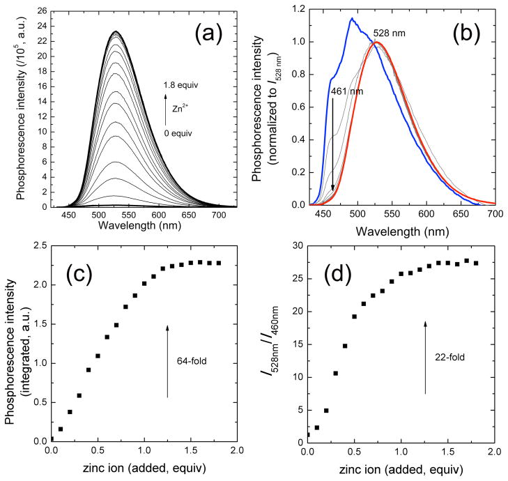
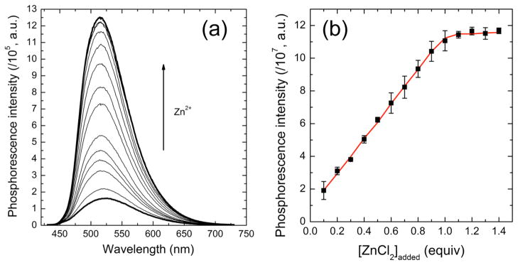
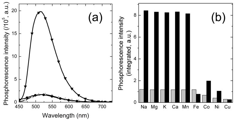
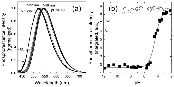
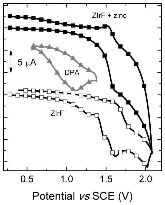
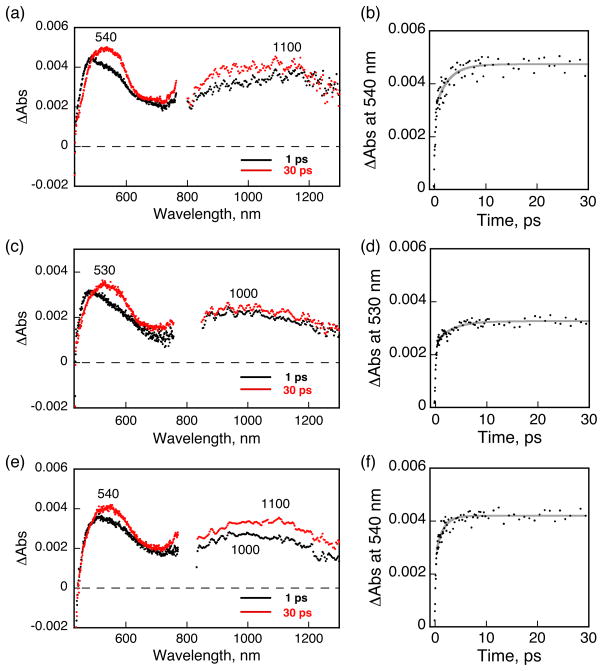
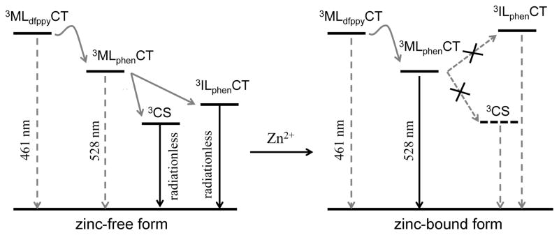
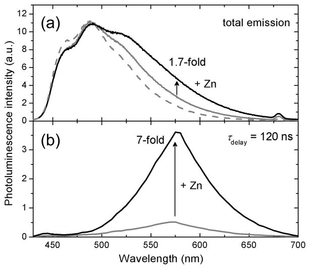
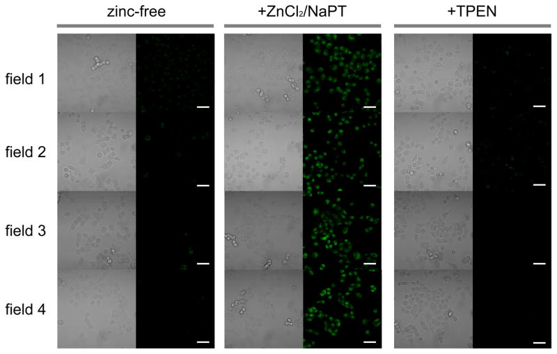
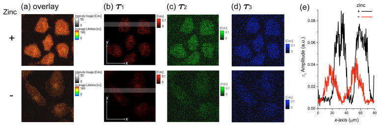
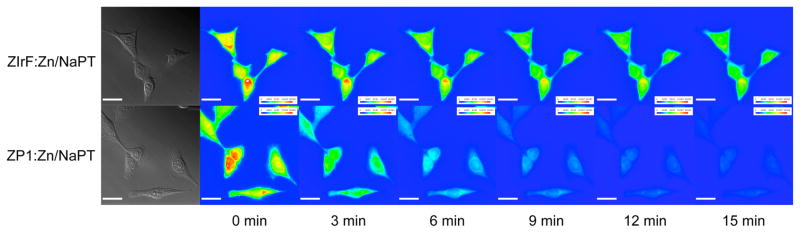
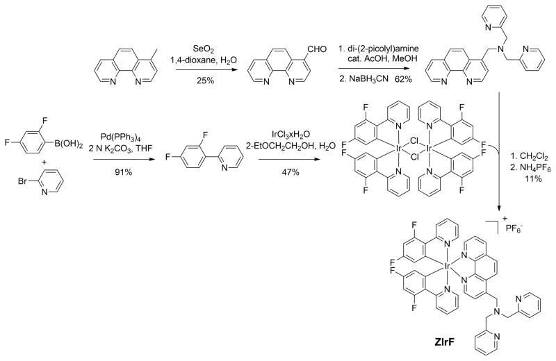
A new phosphorescent zinc sensor (ZIrF) was constructed, based on an Ir(III) complex bearing two 2-(2,4-difluorophenyl)pyridine (dfppy) cyclometalating ligands and a neutral 1,10-phenanthroline (phen) ligand. A zinc-specific di(2-picolyl)amine (DPA) receptor was introduced at the 4-position of the phen ligand via a methylene linker. The cationic Ir(III) complex exhibited dual phosphorescence bands in CH(3)CN solutions originating from blue and yellow emission of the dfppy and phen ligands, respectively. Zinc coordination selectively enhanced the latter, affording a phosphorescence ratiometric response. Electrochemical techniques, quantum chemical calculations, and steady-state and femtosecond spectroscopy were employed to establish a photophysical mechanism for this phosphorescence response. The studies revealed that zinc coordination perturbs nonemissive processes of photoinduced electron transfer and intraligand charge-transfer transition occurring between DPA and phen. ZIrF can detect zinc ions in a reversible and selective manner in buffered solution (pH 7.0, 25 mM PIPES) with K(d) = 11 nM and pK(a) = 4.16. Enhanced signal-to-noise ratios were achieved by time-gated acquisition of long-lived phosphorescence signals. The sensor was applied to image biological free zinc ions in live A549 cells by confocal laser scanning microscopy. A fluorescence lifetime imaging microscope detected an increase in photoluminescence lifetime for zinc-treated A549 cells as compared to controls. ZIrF is the first successful phosphorescent sensor that detects zinc ions in biological samples.


Figures

Figure 1
Figure 1
Calculated geometry and isosurface plot (0.04 e Å−3) of molecular orbitals that participate in the lowest energy triplet transition of ZIrF and IrF, as obtained from Gaussian 03.
Figure 2
Figure 2
(a) Photoluminescence action spectrum of ZIrF (298 K). Inset figure is phosphorescence spectrum at λex = 340 nm. (b) Phosphorescence spectra at various temperatures (λex = 340 nm). Room temperature time-resolved emission spectra (TRES; λex = 342 nm) of ZIrF in the (c) absence and (d) presence of zinc ion (3 equiv). Inset figures are normalized phosphorescence spectra at delay times of 8 and 72 ns. Identical depth scale is employed for (c) and (d). 10 μM ZIrF in air-equilibrated CH3CN solutions were used for measurements.
Figure 3
Figure 3
(a) Change in phosphorescence spectrum with increasing total zinc concentration. (b) Spectra shown in (a) are plotted to have same intensities at 528 nm: Blue line, zinc-free; red line, 1.8 equiv of zinc ion. (c) A titration isotherm plotting phosphorescence intensity as a function of amount of added zinc ion (equiv). (d) Change in phosphorescence intensity ratio at 528 nm vs 460 nm with various amount of added zinc ion. Condition: 10 μM ZIrF in air-equilibrated CH3CN, λex = 340 nm.
Figure 4
Figure 4
(a) Change in phosphorescence spectrum with varying total zinc concentration. (b) Corresponding phosphorescence titration isotherm. The red solid line is a theoretical fit. Refer to text for the theoretical model. Conditions: 10 μM ZIrF in air-equilibrated pH 7.0 buffer (25 mM PIPES), λex = 340 nm.
Figure 5
Figure 5
(a) Phosphorescence spectra of ZIrF showing reversible binding with zinc ions. Empty triangles, zinc-free solution; filled inverted triangles, in the presence of zinc ion (1 equiv); filled circles, after subsequent addition of TPEN (5 equiv). (b) Phosphorescent zinc selectivity of ZIrF. Grey bars, in the presence of metal ions; black bars, after subsequent addition of ZnCl2 (1 equiv). Na+, Mg2+, K+, and Ca2+ ions are 100 equiv. Other divalent metal ions are 1 equiv. Chloride salts were used. Conditions: 10 μM ZIrF in air-equilibrated pH 7.0 buffer (25 mM PIPES), λex = 340 nm.
Figure 6
Figure 6
(a) Change in phosphorescence spectra of ZIrF at decreasing pH from 11.8 to 2.3. (b) pH titration of phosphorescence intensity for zinc-free form (filled squares) and zinc-bound form (4 equiv of ZnCl2; empty circles) of ZIrF. Conditions: 10 μM ZIrF in air-equilibrated milli-Q water containing 100 mM KCl, λex = 340 nm.
Figure 7
Figure 7
Cyclic voltammogram of ZIrF in the absence (empty squares) or presence (filled squares) of Zn(ClO4)2 (5 equiv). The cyclic voltammogram of di(2-picolyl)amine (DPA) is shown for comparison (filled triangles). Conditions: Scan rate = 100 mV/s; 1 mM in Ar-saturated CH3CN containing Bu4NPF6 (0.1 M) supporting electrolyte; Pt wire counter and working electrodes; and the Ag/AgNO3 couple for the reference electrode.
Figure 8
Figure 8
Femtosecond transient absorption spectra measured at 1 and 30 ps delay (left panels) and absorption time profiles (right panels) for (a, b) IrF (0.3 mM), (c, d) Zn-free form (0.3 mM), and (e, f) Zn-bound form of ZIrF (0.3 mM, 1 mM zinc ion). Ar-saturated CH3CN solutions were excited at 420 nm.
Figure 9
Figure 9
Schematic representation of the mechanism for zinc-induced phosphorescence turn-on of ZIrF (MLdfppyCT, metal-to-dfppy ligand charge-transfer transition state; MLphenCT, metal-to-phen ligand charge-transfer transition state; CS, charge-separated state; ILphenCT, intraligand charge-transfer transition state of phen ligand).
Figure 10
Figure 10
Time-gated acquisition of zinc-induced photoluminescence turn-on of ZIrF. (a) Total photoluminescence spectrum of an air-equilibrated pH 7.0 buffer solution (25 mM PIPES) containing ZIrF (10 μM) and Acr+ (2 μM): Solid grey line, total photoluminescence spectrum; dashed grey line, fluorescence spectrum of Acr+ (2 μM); solid black line, after addition of ZnCl2 (5 equiv). (b) Time-gated photoluminescence spectrum acquired after 120 ns delay: Grey line, zinc-free state; black line, after addition of ZnCl2 (5 equiv). Excitation wavelength was 342 nm.
Figure 11
Figure 11
Phosphorescent detection of exogenously supplied intracellular zinc ions in live A549 cells. A549 cells were incubated with ZIrF (5 μM, 30 min; left columns), then with ZnCl2/NaPT (1:1, v/v, 50 μM, 15 min; middle columns), and finally with TPEN (100 μM, 15 min; right columns): Left panels, bright field images; right panels, phosphorescence images. Scale bar corresponds to 50 μm. Identical intensity scale has been applied. Images were acquired from four independent experiments (field 1 4).
Figure 12
Figure 12
(a) Fluorescence lifetime microscope images (80 μm × 80 μm) of fixed A549 cells treated with ZIrF (5 μM, 30 min). Cells in the upper panels were incubated with ZnCl2/NaPT (10 μM, 15 min) prior to ZIrF treatment. (a) Overlay of images (b), (c), and (d): (b) Long-lived component (τ1) image; (c) mid-range component (τ2) image; (d) short-lived component (τ3) image. Refer to Table 3 for the values of τ1, τ2, τ3, and their amplitudes. The same intensity scale is applied as for the lifetime images. (e) Amplitude plots of x-scan for grey regions in image (b).
Figure 13
Figure 13
Comparison of photostability of ZIrF and ZP1. HeLa cells were incubated with Zn/NaPT, fixed, and then treated with either ZIrF or ZP1. Photoluminescence microscope images were taken every three min as the cells were illuminated by the excitation beam.
Scheme 1
Scheme 1
Synthesis of the phosphorescent zinc sensor, ZIrF
Chart 1
Chart 1
Structures of the Phosphorescent Zinc Sensor ZIrF and the Reference Probe IrF

References

    1. Burdette SC, Lippard SJ. Coord Chem Rev. 2001;216–217:333–361.
    1. Palmer AE, Franz KJ. Chem Rev. 2009;109:4533–4535. - PubMed
    1. Que EL, Domaille DW, Chang CJ. Chem Rev. 2008;108:1517–1549. - PubMed
    1. da Silva JJRF, Williams RJP. The Biological Chemistry of Elements: The Inorganic Chemistry of Life. 2. Oxford UP; New York: 2001.
    1. Frederickson CJ. Int Rev Neurobiol. 1989;31:145–238. - PubMed

Publication types