. 2011 Nov;16(11):1071-80.
doi: 10.1111/j.1365-2443.2011.01552.x.

Structural basis for improved efficacy of therapeutic antibodies on defucosylation of their Fc glycans

Affiliations
Free PMC article

Structural basis for improved efficacy of therapeutic antibodies on defucosylation of their Fc glycans

Tsunehiro Mizushima et al. Genes Cells. 2011 Nov.
Free PMC article

Abstract

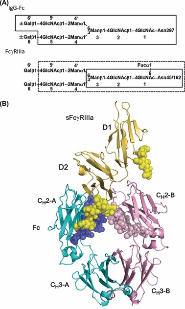
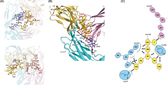
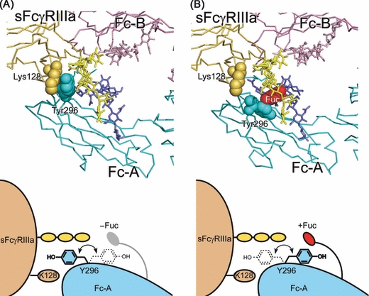
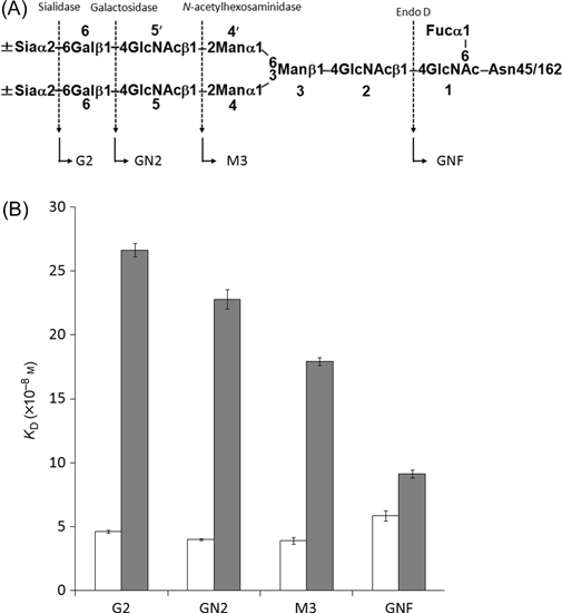
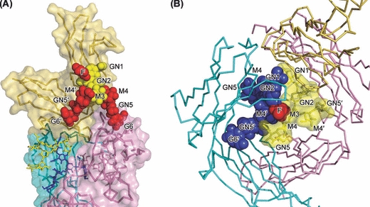
Removal of the fucose residue from the N-glycans of the Fc portion of immunoglobulin G (IgG) results in a dramatic enhancement of antibody-dependent cellular cytotoxicity (ADCC) through improved affinity for Fcγ receptor IIIa (FcγRIIIa). Here, we present the 2.2-Å structure of the complex formed between nonfucosylated IgG1-Fc and a soluble form of FcγRIIIa (sFcγRIIIa) with two N-glycosylation sites. The crystal structure shows that one of the two N-glycans of sFcγRIIIa mediates the interaction with nonfucosylated Fc, thereby stabilizing the complex. However, fucosylation of the Fc N-glycans inhibits this interaction, because of steric hindrance, and furthermore, negatively affects the dynamics of the receptor binding site. Our results offer a structural basis for improvement in ADCC of therapeutic antibodies by defucosylation.


Figures

Figure 1
Figure 1
Structure of nonfucosylated Fc complexed with bis-glycosylated soluble form of Fcγ receptor IIIa (sFcγRIIIa). (A) N-glycans expressed on immunoglobulin G (IgG)-Fc and sFcγRIIIa used for crystallization. Sugar residues that gave interpretable electron densities are surrounded by solid polygons in Fc-Asn-297 N-glycans and sFcγRIIIa-Asn-162 N-glycan, whereas those in the sFcγRIIIa-Asn-162 N-glycan are indicated by a dashed box. (B) Overall view of the complex. Chains A and B of Fc are cyan and pink, respectively, and sFcγRIIIa is yellow. Carbohydrates are shown in sphere representation. F, Fuc, fucose; G, Gal, galactose; GN, GlcNAc, N-acetylglucosamine; M, Man, mannose.
Figure 2
Figure 2
Binding of nonfucosylated Fc to soluble form of Fcγ receptor IIIa (sFcγRIIIa) is mediated by carbohydrate–protein and carbohydrate–carbohydrate interactions. (A) Close-up view of the interface between sFcγRIIIa-Asn162 N-glycan and the Fc N-glycans [in chain A (upper panel) and chain B (lower panel)]. Hydrogen bonds are represented by dotted lines. (B) Close-up view of the interface between the sFcγRIIIa-Asn162 N-glycan (yellow) and Fc chain A (cyan) (C) Schematic representation of the carbohydrate–carbohydrate interactions.
Figure 3
Figure 3
Alternative modes of Fc–soluble form of Fcγ receptor IIIa (sFcγRIIIa) interaction with different orientations of the Tyr-296 aromatic ring depending on Fc fucosylation. (A) The present crystal structure, in which the aromatic ring of Tyr-296 of nonfucosylated Fc (chain A) is flipped out and sandwiched between Man-4 of the Asn-162 glycan and Lys-128 of sFcγRIIIa. (B) 3D model of sFcγRIIIa bound to fucosylated Fc, in which the tyrosine ring does not make contact with the receptor, but makes intramolecular contact with the core fucose residue. The model is based on the crystal structure of the complex between fucosylated Fc and nonglycosylated sFcγRIIIb (1E4K) with substitution of the receptor molecule by bis-glycosylated sFcγRIIIb in the present crystal structure by superposing its D2 domains. In this model, the core fucose residue was attached to GlcNAc-1 by the α1–6 glycosidic linkage in the N-glycans in Fc chain A by superimposing the crystal structure of the fucosylated IgG1-Fc (3AVE). Chains A and B of Fc are cyan and pink, respectively, and sFcγRIIIa is yellow.
Figure 4
Figure 4
Glycoform-dependent interactions between human immunoglobulin G1 (IgG1) and soluble form of Fcγ receptor IIIa (sFcγRIIIa). (A) Carbohydrate sequence of sFcγRIIIa used in the present study. Dotted arrows indicate cleavage sites of glycosidases. The resulting glycoforms are indicated by arrows. (B) White and gray bars represent KD values of the binding of sFcγRIIIa glycoforms to nonfucosylated and fucosylated IgG glycoproteins, respectively. These values were calculated based on SPR data.
Figure 5
Figure 5
Potential steric hindrance effects in interactions between (A) CH2 domain of Fc and soluble form of Fcγ receptor IIIa (sFcγRIIIa)-Asn-45 N-glycan and (B) fucosylated N-glycan of Fc chain A and sFcγRIIIa-Asn-162 N-glycan. The sugar residues of the Asn-45 N-glycan gave no interpretable electron density, and the core fucose residues of the Fc N-glycan were modeled and displayed in red. The modeling was based on the conformations of the N-glycan of Fc chain A (for the lactosamine branches in A) and the Asn-162 N-glycan (for the fucose residues in A and B).

Similar articles

Cited by

References

    1. Behring E, Kitasato S. Ûber das zustandekommen der Díeptherid-Immunitat und der Tetanus-Immunitat bei Tieren. Dtsch. Med. Wochenschr. 1890;16:1113–1114. - PubMed
    1. Burton DR, Woof JM. Human antibody effector function. Adv. Immunol. 1992;51:1–84. - PubMed
    1. CCP4 The CCP4 suite: programs for protein crystallography. Acta Crystallogr. D Biol. Crystallogr. 1994;50:760–763. - PubMed
    1. DeLano WL. The PyMOL Molecular Graphics System. San Carlos, CA: DeLano Scientific; 2002.
    1. Emsley P, Cowtan K. Coot: model-building tools for molecular graphics. Acta Crystallogr. D Biol. Crystallogr. 2004;60:2126–2132. - PubMed

Publication types

MeSH terms

Associated data