Review
. 2011 Dec;46(6):507-26.
doi: 10.3109/10409238.2011.619164. Epub 2011 Oct 25.

Epigenetic virtues of chromodomains

Affiliations
Review

Epigenetic virtues of chromodomains

Bartlomiej J Blus et al. Crit Rev Biochem Mol Biol. 2011 Dec.

Abstract

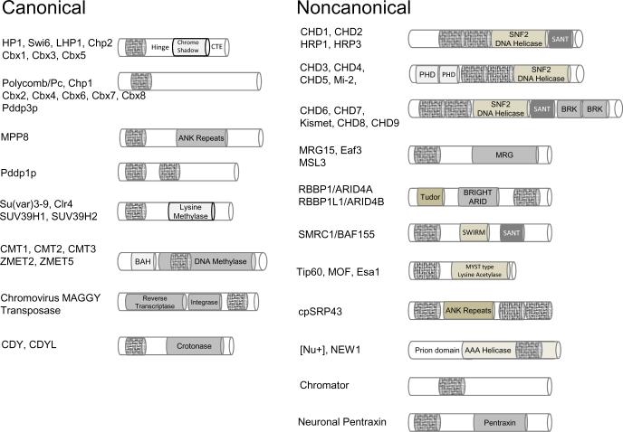
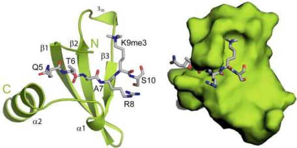
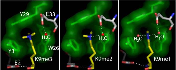
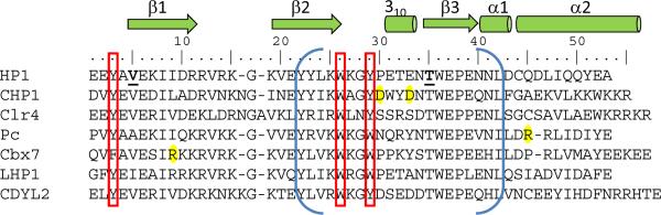
The chromatin organization modifier domain (chromodomain) was first identified as a motif associated with chromatin silencing in Drosophila. There is growing evidence that chromodomains are evolutionary conserved across different eukaryotic species to control diverse aspects of epigenetic regulation. Although originally reported as histone H3 methyllysine readers, the chromodomain functions have now expanded to recognition of other histone and non-histone partners as well as interaction with nucleic acids. Chromodomain binding to a diverse group of targets is mediated by a conserved substructure called the chromobox homology region. This motif can be used to predict methyllysine binding and distinguish chromodomains from related Tudor "Royal" family members. In this review, we discuss and classify various chromodomains according to their context, structure and the mechanism of target recognition.


Figures

Figure 1
Figure 1
Diverse chromodomains recognize methyl-lysine marks in H3 and H4 tails. a) Composition of chromodomain proteins, and their classification into canonical and noncanonical polypeptides. Chromodomain module is represented with a hatched rod. (b) Schematic of the absolutely conserved histone H3 and H4 tails and their locations in the nucleosome core particle. Sites of modifications with methylation of lysine (blue star), methylation of arginine (green star), phosphorylation of threonine (red square) and acetylation of lysines (orange circle) are labeled in histone tail sequences.
Figure 1
Figure 1
Diverse chromodomains recognize methyl-lysine marks in H3 and H4 tails. a) Composition of chromodomain proteins, and their classification into canonical and noncanonical polypeptides. Chromodomain module is represented with a hatched rod. (b) Schematic of the absolutely conserved histone H3 and H4 tails and their locations in the nucleosome core particle. Sites of modifications with methylation of lysine (blue star), methylation of arginine (green star), phosphorylation of threonine (red square) and acetylation of lysines (orange circle) are labeled in histone tail sequences.
Figure 2
Figure 2
Structural properties of canonical chromodomains. a) Ribbon and surface representation of the HP1 chromodomain (green) bound to the H3K9me3 peptide (grey). (b) Differentially methylated peptides (yellow) form the similar cation-π interactions with the aromatic cage of the HP1 chromodomain (green). The free hydrogens in mono- and dimethyllysine form water-mediated hydrogen bonds (dotted lines) with the chromodomain. c) Sequence alignment of HP1 chromodomain with other well-studied canonical chromodomains. The red bars depict the aromatic residues of the cage. The blue brackets correspond to the chromobox homology motif. In bold is the threonine that resides in the Casein Kinase II consensus sequence. Highlighted in yellow are the residues that uniquely contribute to structure of complexes.
Figure 2
Figure 2
Structural properties of canonical chromodomains. a) Ribbon and surface representation of the HP1 chromodomain (green) bound to the H3K9me3 peptide (grey). (b) Differentially methylated peptides (yellow) form the similar cation-π interactions with the aromatic cage of the HP1 chromodomain (green). The free hydrogens in mono- and dimethyllysine form water-mediated hydrogen bonds (dotted lines) with the chromodomain. c) Sequence alignment of HP1 chromodomain with other well-studied canonical chromodomains. The red bars depict the aromatic residues of the cage. The blue brackets correspond to the chromobox homology motif. In bold is the threonine that resides in the Casein Kinase II consensus sequence. Highlighted in yellow are the residues that uniquely contribute to structure of complexes.
Figure 3
Figure 3
Sequence inserts in noncanonical chromodomains modulate binding activity. The canonical architecture of chromodomains is displayed in green, inserts are in brown, aromatic cage residues are drawn in ball and stick, and peptides are in gray. a) The double chromodomains of human CHD1 linked by a novel helix-turn-helix (gray). (b) The solution structure of the yeast Eaf3 chromodomain with its C-terminal extension containing a methyllysine analog mimicking the binding of the H3K36 methyl-mark to its aromatic cage. (c) The MSL3 chromodomain in complex with a DNA duplex promoting its selectivity for the H4K20me1 peptide. The peptide sits on the DNA minor groove, with its methyllysine inserted in the four-residue aromatic cage.
Figure 4
Figure 4
Noncanonical chromodomains that lack an aromatic cage. The color-coding is similar to that in Figure 3. (a) The Drosophila MOF chromodomain also called a chromobarrel domain. (b) Esa1 chromodomain, also called a knotted-tudor domain. (c) The human chromo shadow dimer in complex with the EMSY peptide. (d) Chromodomain 1 of cpSRP43. (e) The structurally-observed chromodomain-like domain in the eEF3.
Figure 5
Figure 5
The chromobox homology motif is a toolkit for distinguishing the tudor clan modules from the chromodomain. a) A sequence alignment of the chromobox from a selection of chromodomains and related domains: chromo shadow domain, tudor, MBT, PWWP and Agenet. b–d) Ribbon diagram show superposition of the relevant regions of homology and point to key aromatic residues. The superposition of the Drosophila HP1 chromobox with related regions from other domains produced the following rmsd over the backbone residues: 1.21 Å with MBT, 0.44 Å with MSL3, 1.5 Å with 53BP1 Tudor, 2.7 Å with FRMP Agenet, 0.96 Å with chromo shadow and 1.45 Å with PWWP.

References

    1. AASLAND R, STEWART AF. The chromo shadow domain, a second chromo domain in heterochromatin-binding protein 1, HP1. Nucleic Acids Res. 1995;23:3168–73. - PMC - PubMed
    1. AKHTAR A, BECKER PB. Activation of transcription through histone H4 acetylation by MOF, an acetyltransferase essential for dosage compensation in Drosophila. Mol Cell. 2000;5:367–75. - PubMed
    1. AKHTAR A, ZINK D, BECKER PB. Chromodomains are protein-RNA interaction modules. Nature. 2000;407:405–9. - PubMed
    1. ALEKSEYENKO AA, PENG S, LARSCHAN E, GORCHAKOV AA, LEE OK, KHARCHENKO P, MCGRATH SD, WANG CI, MARDIS ER, PARK PJ, KURODA MI. A sequence motif within chromatin entry sites directs MSL establishment on the Drosophila X chromosome. Cell. 2008;134:599–609. - PMC - PubMed
    1. ALLIS CD, BERGER SL, COTE J, DENT S, JENUWIEN T, KOUZARIDES T, PILLUS L, REINBERG D, SHI Y, SHIEKHATTAR R, SHILATIFARD A, WORKMAN J, ZHANG Y. New nomenclature for chromatin-modifying enzymes. Cell. 2007;131:633–6. - PubMed

Publication types

LinkOut - more resources