. 2012 Jan;233(1):221-32.
doi: 10.1016/j.expneurol.2011.10.005. Epub 2011 Oct 14.

The transcription factor Sox11 promotes nerve regeneration through activation of the regeneration-associated gene Sprr1a

Affiliations

The transcription factor Sox11 promotes nerve regeneration through activation of the regeneration-associated gene Sprr1a

Xiaotang Jing et al. Exp Neurol. 2012 Jan.

Abstract

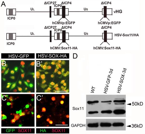
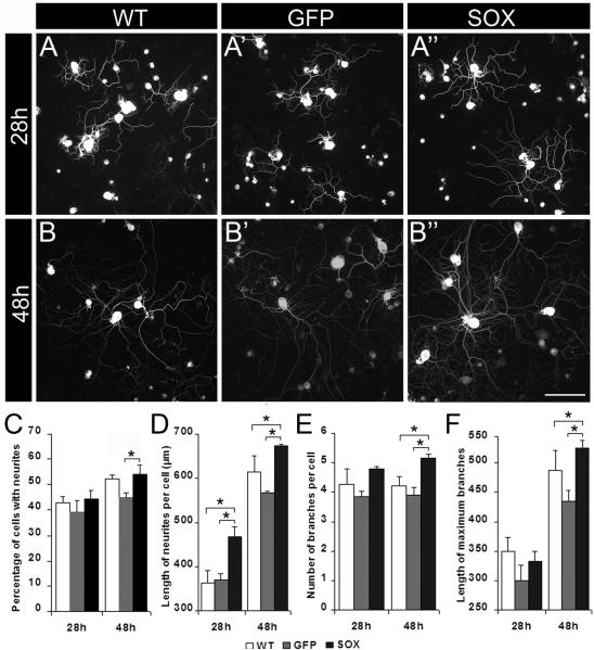
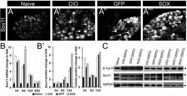
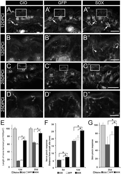
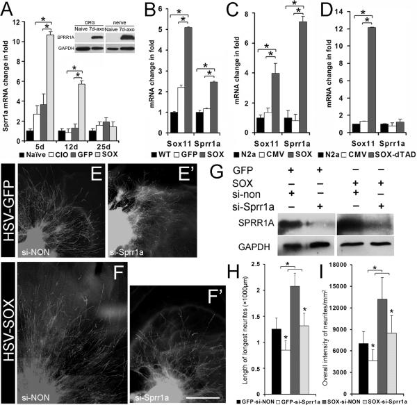
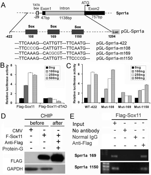
Factors that enhance the intrinsic growth potential of adult neurons are key players in the successful repair and regeneration of neurons following injury. Injury-induced activation of transcription factors has a central role in this process because they regulate expression of regeneration-associated genes. Sox11 is a developmentally expressed transcription factor that is significantly induced in adult neurons in response to injury. Its function in injured neurons is however undefined. Here, we report studies that use herpes simplex virus (HSV)-vector-mediated expression of Sox11 in adult sensory neurons to assess the effect of Sox11 overexpression on neuron regeneration. Cultured mouse dorsal root ganglia (DRG) neurons transfected with HSV-Sox11 exhibited increased neurite elongation and branching relative to naïve and HSV-vector control treated neurons. Neurons from mice injected in foot skin with HSV-Sox11 exhibited accelerated regeneration of crushed saphenous nerves as indicated by faster regrowth of axons and nerve fibers to the skin, increased myelin thickness and faster return of nerve and skin sensitivity. Downstream targets of HSV-Sox11 were examined by analyzing changes in gene expression of known regeneration-associated genes. This analysis in combination with mutational and chromatin immunoprecipitation assays indicates that the ability of Sox11 to accelerate in vivo nerve regeneration is dependent on its transcriptional activation of the regeneration-associated gene, small proline rich protein 1a (Sprr1a). This finding reveals a new functional linkage between Sox11 and Sprr1a in adult peripheral neuron regeneration.


Figures

Figure 1
Figure 1. HSV mediated gene transfer of Sox11 in vitro
A. Genomic structure of HSV vectors used for GFP and Sox11 gene transfer. The control vHG vector contains deletions (Δ) of ICP4 and ICP27 and a hCMVp:eGFP cassette. The hCMV:Sox11 or hCMV:Sox11-HA cassettes were also targeted to the two ICP4 loci. B-C’. GFP fluorescence and antibodies to Sox11 (red) or HA (green) were used to detect HSV vector expression in Neuro2A cells (B-B’) and DRG neurons (C-C’) at 24h after vector addition. Note increase in Sox11 labeling intensity in HSV-Sox11-HA treated cultures (red labeling in B’ and C’) relative to HSV-GFP (B and C) treated cultures. D. Immunoblots assessed Sox11 protein at 3d after HSV-Sox11-HA infection in cultured DRG neurons. Bands just above 50 kD identify Sox11 since they align with bands present in Neuro2A cells transfected with CMV-Sox11 plasmids (not shown). The identity of the major lower band is unknown at this time. Also note that HSV-driven expression of GFP in cultured neurons causes a reduction in Sox11 protein level. This effect was not seen in vivo (see Fig. 3C) and may reflect the high level of GFP protein. Scale bars=50μm. GAPDH used as loading control was unchanged.
Figure 2
Figure 2. HSV mediated gene transfer of Sox11 promotes neurite extension and branching in vitro
Representative images show DRG neurons that were untreated (A, B), infected with HSV-GFP (A’, B’) or with HSV-Sox11 vectors (A’’, B’’). Analysis of neurons labeled with anti-β-tubulin III showed HSV-Sox11 increased average neurite length at 28h and 48h post treatment (D, P<0.01). At 48h the number of branches per cell (E) and the length of the longest branch (F) also increased relative to untreated control groups (P<0.05). At 48h, HSV-Sox11 treated neurons had more neurites but only relative to HSV-GFP controls (C, P<0.01); this may reflect a GFP-specific inhibition in cultured neurons. No other difference between WT and HSV-GFP cultures was significant. Data is collected from 3 independent experiments with over 600 cells analyzed per group. Scale bar in B” = 100 μm.
Figure 3
Figure 3. HSV-Sox11 increases Sox11 expression in injured peripheral neurons
DRG from naïve mice and mice with saphenous nerve injury were collected at 5DPCI and immunolabeled with anti-Sox11 (A-A’’’). Increased immunoreactivity occurs in CIO, HSV-GFP and HSV-Sox11 sections relative to naïve sections. Arrowheads indicate smaller neurons that are Sox11-positive. B. Sox11 mRNA increased in all treatment groups relative to naïve with the greatest change in HSV-Sox11 DRG at 5d post injury (P<0.05, n=6). B’-C. Increased levels of Sox11 protein shown by western blot analysis of L2/L3 DRG collected at 3, 5 and 12 DPCI. Sox11 increases in HSV-Sox11 treated mice relative to naïve, CIO or GFP controls. β-TubIII expression was unchanged across treatment groups (B’). Scale bar in A”’ = 100 μm.
Figure 4
Figure 4. Histomorphometric analysis of saphenous nerves
Light level micrographs of toluidine blue stained transverse semi-thin sections at 3 mm distal to the injury were used to determine the number of myelinated fibers and myelin thickness. Representative images of nerves collected at 5 (A-A’’), 12 (B-B’’) and 25 DPCI (C-C’’) from CIO (A, B, C), HSV-GFP (A’, B’, C’) and HSV-Sox11 groups (A’’, B’’, C’’) are shown. Naïve littermate (E) is included for comparison. At 5d crushed nerves were swollen with degenerating axons with collapsed myelin. A significant increase in both the number of myelinated profiles (D) and average area of myelin (D’) occurs by 25d in nerves of HSV-Sox11 treated animals. n = 4 per group, * P<0.01, determined by two-tailed Student's t-test. Scale bar in E = 20μm.
Figure 5
Figure 5. HSV-Sox11 enhances nerve regeneration to the skin and functional recovery
Saphenous nerves were crushed at 3d post HSV inoculation and skin overlying the saphenous innervation field (see Fig. A1) was analyzed. Shown are representative images of sections stained with PGP9.5 taken at low (A-A”, C-C”) and higher (B-B”, D-D”) magnification at 12 (A-B”) and 25 (C-D’’) DPCI. Boxed areas indicate region of magnification. Few dermal fibers (arrows) and virtually no epidermal fibers were visible at 12 DPCI in CIO and HSV-GFP samples (A, A’). However, skin of HSV-Sox injected animals showed numerous fibers in the dermis (arrows, A’’) and approaching the epidermis (arrowheads, B”). At 25 DPCI, epidermal fibers were present in all treatment groups (C-D’’). E. Overall length of PGP9.5-positive fibers relative to epidermal length at 12 and 25 DPCI is greatest in HSV-Sox11 injected skin. F. Nerve responses to pinch assessed at 5 and 12 DPCI showed greater responses in Sox11 treated animals. G. A skin pinch test also showed HSV-Sox11 treatment accelerated functional recovery of skin in the saphenous nerve field at 25 DPCI (n=6 per group). Scale bar in A” and C” = 50 μm, in B” and D” = 15μm; n=6 in F and G. * P<0.01.
Figure 6
Figure 6. Sprr1a transcription is required for stimulation of axon growth by Sox11
A. Saphenous nerve crush increases Sprr1a mRNA in ganglia of HSV-Sox11 treated mice at 5 and 12 DPCI (n=6 at 5DPCI, all other days n=4, asterisks indicate P<0.001). Inset: Western blot shows rise in Sprr1a protein in DRG and sciatic nerve at 7d post sciatic axotomy as previously reported (Bonilla, et al., 2002). B. HSV-Sox11 increases Sprr1a expression in cultured DRG neurons relative to WT and HSV-GFP control groups. Measures were made at 6d postinfection (cultures derived from n=3 mice, P<0.01). C. Neuro2A cells transfected with pCMV-Sox11 plasmid increase Sprr1a mRNA at 24h post transfection (n=3, P<0.01). D. Sprr1a gene activation does not occur in Neuro2A cells transfected with a mutant Sox11 that lacks the transactivation domain (Sox-11FΔTAD) (n=3, asterisks indicate P<0.01). E-I. Sprr1a transcription is required for Sox11 stimulation of axon growth. HSV-GFP or HSV-Sox11 treated DRG explants (E-F’) or dissociated cell cultures (G) were transfected with non-targeting (si-NON; E, F) or Sprr1a targeted siRNAs (si-Sprr1a; E’, F’). HSV-Sox11 explants treated with non-targeting siRNA (F) showed enhanced neurite growth relative to HSV-GFP controls (E). Treatment with siRNA to Sprr1a blocked the Sox11 effect (F’). Western blots in G show effective Sprr1a knockdown in DRG cultures treated with HSV-GFP or HSV-Sox11. Quantification of neurite lengths and intensity of TuJ1-positive neurite labeling are plotted in H and I. * P<0.05.
Figure 7
Figure 7. Sox11 regulates transcription of Sprr1a
A. Diagram of the Sprr1a promoter/intron construct used in this study. Small grey boxes indicate predicted Sox binding sites at nt108, nt169 and nt1150 with the start of numbering at the transcription start site. Sequences of core nucleotides of wildtype and mutated Sox sites (grey lettering) are shown below. B. pGL-Sprr1a-luciferase plasmids were transfected into Neuro2A cells in combination with 0, 100, 250, 500ng of empty CMV vector, pCMV-Sox11F or pCMVSox-11FΔTAD. Three independent experiments were performed in triplicate and normalized to the CMV empty vector whose value was set to 1. C. pGL2-Sprr1a-422 activity is suppressed following mutation of Sox-binding sites at nt169 and nt1150 but not nt108. D. Western blot shows enrichment of Sox-11F protein in ChIP samples immunoprecipitated by anti-FLAG antibody. E. ChIP assay shows Sox11 binding to endogenous Sprr1a at nt169 and nt1150 sites. Chromatin extracts from Neuro2A cells transiently expressing Sox-11F were precipitated with no antibody, with non-immune mouse IgG or with FLAG antibody. PCR products from immunoprecipitated chromatin and starting input are shown on ethidium stained agarose gel.

Similar articles

Cited by

References

    1. Abe N, Cavalli V. Nerve injury signaling. Curr Opin Neurobiol. 2008;18:276–283. - PMC - PubMed
    1. Badis G, Berger MF, Philippakis AA, Talukder S, Gehrke AR, Jaeger SA, Chan ET, Metzler G, Vedenko A, Chen X, Kuznetsov H, Wang CF, Coburn D, Newburger DE, Morris Q, Hughes TR, Bulyk ML. Diversity and complexity in DNA recognition by transcription factors. Science. 2009;324:1720–1723. - PMC - PubMed
    1. Bergsland M, Werme M, Malewicz M, Perlmann T, Muhr J. The establishment of neuronal properties is controlled by Sox4 and Sox11. Genes Dev. 2006;20:3475–3486. - PMC - PubMed
    1. Bhattaram P, Penzo-Mendez A, Sock E, Colmenares C, Kaneko KJ, Vassilev A, Depamphilis ML, Wegner M, Lefebvre V. Organogenesis relies on SoxC transcription factors for the survival of neural and mesenchymal progenitors. Nat Commun. 2010;1:9. - PMC - PubMed
    1. Bonilla IE, Tanabe K, Strittmatter SM. Small proline-rich repeat protein 1A is expressed by axotomized neurons and promotes axonal outgrowth. J Neurosci. 2002;22:1303–1315. - PMC - PubMed

Publication types

MeSH terms