. 2012 Feb;26(2):917-26.
doi: 10.1096/fj.11-195883. Epub 2011 Oct 24.

Nicotinic acetylcholine receptors in dorsal root ganglion neurons include the α6β4* subtype

Affiliations

Nicotinic acetylcholine receptors in dorsal root ganglion neurons include the α6β4* subtype

Arik J Hone et al. FASEB J. 2012 Feb.

Abstract

The α6-containing nicotinic acetylcholine receptors (nAChRs) have recently been implicated in diseases of the central nervous system (CNS), including Parkinson's disease and substance abuse. In contrast, little is known about the role of α6* nAChRs in the peripheral nervous system (where the asterisk denotes the possible presence of additional subunits). Dorsal root ganglia (DRG) neurons are known to express nAChRs with a pharmacology consistent with an α7, α3β4*, and α4β2* composition. Here we present evidence that DRG neurons also express α6* nAChRs. We used RT-PCR to show the presence of α6 subunit transcripts and patch-clamp electrophysiology together with subtype-selective α-conotoxins to pharmacologically characterize the nAChRs in rat DRG neurons. α-Conotoxin BuIA (500 nM) blocked acetylcholine-gated currents (I(ACh)) by 90.3 ± 3.0%; the recovery from blockade was very slow, indicating a predominance of α(x)β4* nAChRs. Perfusion with either 300 nM BuIA[T5A;P6O] or 200 nM MII[E11A], α-conotoxins that target the α6β4* subtype, blocked I(ACh) by 49.3 ± 5 and 46.7 ± 8%, respectively. In these neurons, I(ACh) was relatively insensitive to 200 nM ArIB[V11L;V16D] (9.4±2.0% blockade) or 500 nM PnIA (23.0±4% blockade), α-conotoxins that target α7 and α3β2*/α6β2* nAChRs, respectively. We conclude that α6β4* nAChRs are among the subtypes expressed by DRG, and to our knowledge, this is the first demonstration of α6β4* in neurons outside the CNS.


Figures
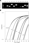
Figure 1.
Figure 1.
RT-PCR analysis of nAChR subunit transcripts. A) Endpoint RT-PCR products were detected for nAChR subunits α3–α7, α10, and β2–β4 and visualized using ethidium bromide fluorescence. Reactions for each primer set were performed in the absence of template (H2O only) as controls and were negative (data not shown). B) Real-time RT-PCR analysis confirms the presence of α3–α7 and α10 nAChR subunit transcripts; transcripts for α2 and α9 were not detected or were below the threshold level of detection at cycle number 40 (data not shown).
Figure 2.
Figure 2.
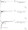
Whole-cell voltage-clamp electrophysiology of DRG neurons. Neurons were held at −80 mV and stimulated with 1 mM ACh for 100 ms every 2 min. Under these conditions, 2 broad types of responses were observed. A) Category I responses were characterized by rapid activation and desensitization kinetics. B) Category II responses were characterized by slower kinetics relative to category I responses.
Figure 3.
Figure 3.
Pharmacological evaluation of category I and II response types. A, B) Category I responses are blocked by α7-selective nAChR antagonists (A); blockade was observed using ArIB[V11L;V16D] (200 nM), and the response slowly recovered during washout (B). C, D) α-Cbtx (100 nM) also blocked category I responses (C); there was very little recovery from blockade even after 20 min of wash (D). E) Category II responses are mainly mediated by heteromeric nAChRs. These responses were relatively insensitive to ArIB[V11L;V16D] (200 nM) but were substantially blocked by BuIA (500 nM). F) In the continued presence of ArIB[V11L;V16D] (200 nM), recovery from blockade by BuIA was very slow; there was little recovery after a 12 min wash. c, control response before application of toxins.
Figure 4.
Figure 4.
Slow kinetics of blockade and recovery from blockade category II responses by BuIA indicate a predominance of αxβ4 nAChRs. A) Superimposed trace recordings of ACh (100 ms pulse every 2 min) responses from a neuron before and during a 20-min perfusion with BuIA (500 nM). All solutions contained ArIB[V11L;V16D] (200 nM) to inhibit any α7 nAChRs. B) Single exponential fit of data showing the observed rate of blockade by BuIA (500 nM); t1/2 = 3.9 min (95% CI: 2.9–5.9). Error bars = se for 6 neurons. C, D) To determine recovery from BuIA blockade, neurons were simultaneously perfused with ArIB[V11L;V16D] (200 nM) and BuIA (5 μM) for 6 min (C), and ACh responses were monitored for recovery during washout of BuIA (in the continued presence of ArIB[V11L;V16D] to keep α7 nAChRs inhibited); there was little recovery after a 20 min wash (D). E) Blockade of α4β2* by DHβE. Near complete blockade of the response was observed by simultaneous perfusion with a combination of BuIA (500 nM), ArIB[V11L;V16D] (200 nM), and DHβE (1 μM). Additional blockade by DHβE is consistent with the presence of α4β2* nAChRs. For comparison, see panels A–C and Fig. 3E. c, control response before perfusion with toxin solution. Panels A, C, E show results from single neurons; see Results for averages of multiple neurons.
Figure 5.
Figure 5.
α-Ctx PnIA blocks α3β2 and α6/α3β2β3 but not α3β4 or α6/α3β4 nAChRs in Xenopus oocytes. A) Blockade of α6/α3β2β3 was determined by perfusing the oocyte with solutions containing increasing concentrations of PnIA and yielded a value of IC50 = 10.9 nM (95% CI: 7.9–14.9), nH = 1.0 (95% CI: 0.7–1.4). Error bars = se for 5 individual experiments. B) Blockade of α6/α3β2β3 by PnIA (500 nM). C–E) PnIA (500 nM) also blocks α3β2 nAChRs (C) but neither α6/α3β4 nAChRs (D) nor α3β4 nAChRs (E). Panels B-E show responses in a single oocyte;see Results for averages of multiple oocytes. /–/ indicates a 5-min exposure to PnIA.
Figure 6.
Figure 6.
Effect of PnIA on category II responses in DRG neurons. PnIA blocks a portion of category II responses. A) Neurons were perfused with ArIB[V11L;V16D] (200 nM) followed by PnIA (500 nM). B) A cocktail of antagonists consisting of ArIB[V11L;V16D] (200 nM), PnIA (500 nM), and DHβE (1 μM), to block α7, α3β2*/α6β2*, and α4β2* nAChRs, respectively, blocked a portion of the response similar to that observed in panel A. c, control response before perfusion with toxins.
Figure 7.
Figure 7.
Category II responses are blocked by α-Ctxs that target α6β4* nAChRs. A panel of nAChR subtype-selective α-Ctxs, listed in Table 1, was used to isolate and block α6β4* nAChRs in neurons with category II responses. A) After perfusion with the cocktail, the α6β4 nAChR antagonist BuIA[T5A;P6O] (300 nM) reversibly blocked a substantial portion of the remaining response. B) Sequential and cumulative application of the cocktail, followed by BuIA[T5A;P6O] (300 nM), then AuIB (10 μM) to block α3β4* nAChRs. C) Omission of DHβE from the cocktail, followed by perfusion with 100 nM, then 300 nM BuIA[T5A;P6O]. D) Following the same protocol as in panel B, perfusion of the cocktail, followed by MII[E11A] (200 nM) to block α6β4* nAChRs, and then AuIB (10 μM). Bottom graphs show time course of blockade by the toxins; bars labeled a–c indicate perfusion duration for each toxin solution and correlate with same labels in trace recordings (top panels). Results are from single neurons; see Results for averages of multiple neurons. c = control response before application of toxin.

References

    1. Van Boxem K., van Bilsen J., de Meij N., Herrler A., Kessels F., Van Zundert J., van Kleef M. (2010) Pulsed radiofrequency treatment adjacent to the lumbar dorsal root ganglion for the management of lumbosacral radicular syndrome: a clinical audit. Pain Med. 12, 1322–1330 - PubMed
    1. Van Zundert J., Patijn J., Kessels A., Lame I., van Suijlekom H., van Kleef M. (2007) Pulsed radiofrequency adjacent to the cervical dorsal root ganglion in chronic cervical radicular pain: a double blind sham controlled randomized clinical trial. Pain 127, 173–182 - PubMed
    1. Simopoulos T. T., Kraemer J., Nagda J. V., Aner M., Bajwa Z. H. (2008) Response to pulsed and continuous radiofrequency lesioning of the dorsal root ganglion and segmental nerves in patients with chronic lumbar radicular pain. Pain Physician 11, 137–144 - PubMed
    1. Genzen J. R., Van Cleve W., McGehee D. S. (2001) Dorsal root ganglion neurons express multiple nicotinic acetylcholine receptor subtypes. J. Neurophysiol. 86, 1773–1782 - PubMed
    1. Dube G. R., Kohlhaas K. L., Rueter L. E., Surowy C. S., Meyer M. D., Briggs C. A. (2005) Loss of functional neuronal nicotinic receptors in dorsal root ganglion neurons in a rat model of neuropathic pain. Neurosci. Lett. 376, 29–34 - PubMed

Publication types

LinkOut - more resources