. 2011 Dec;7(12):1490-9.
doi: 10.4161/auto.7.12.17924.

The induction of autophagy by mechanical stress

Affiliations

The induction of autophagy by mechanical stress

Jason S King et al. Autophagy. 2011 Dec.

Abstract

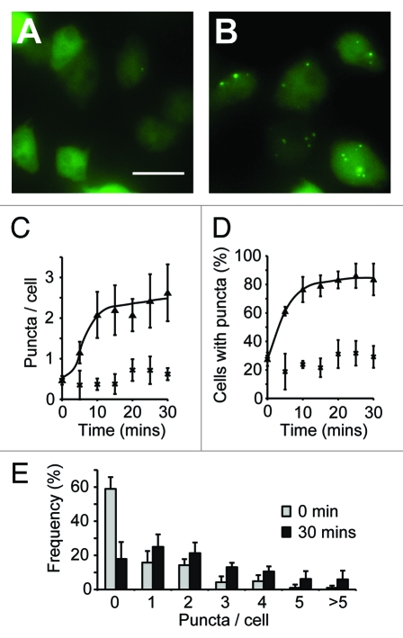
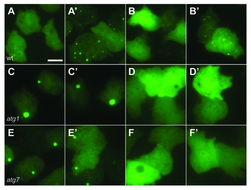
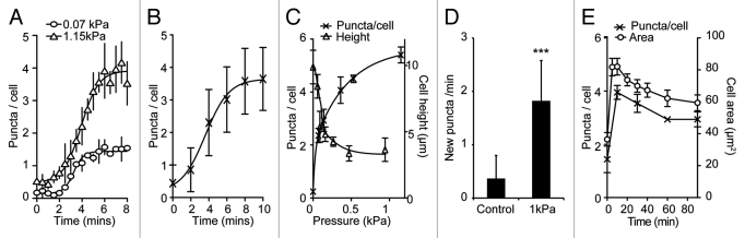
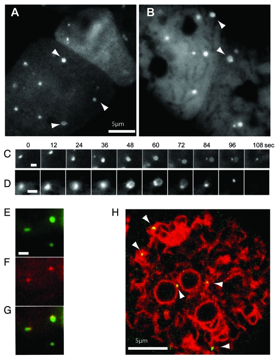
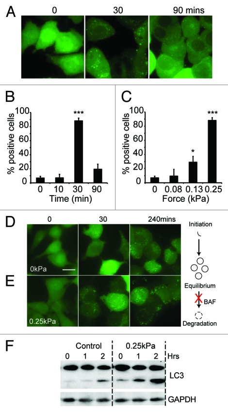
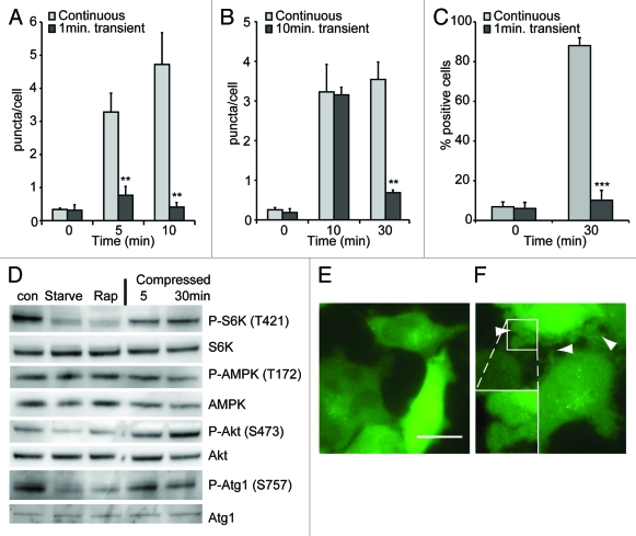
The ability to respond and adapt to changes in the physical environment is a universal and essential cellular property. Here we demonstrated that cells respond to mechanical compressive stress by rapidly inducing autophagosome formation. We measured this response in both Dictyostelium and mammalian cells, indicating that this is an evolutionarily conserved, general response to mechanical stress. In Dictyostelium, the number of autophagosomes increased 20-fold within 10 min of 1 kPa pressure being applied and a similar response was seen in mammalian cells after 30 min. We showed in both cell types that autophagy is highly sensitive to changes in mechanical pressure and the response is graduated, with half-maximal responses at ~0.2 kPa, similar to other mechano-sensitive responses. We further showed that the mechanical induction of autophagy is TOR-independent and transient, lasting until the cells adapt to their new environment and recover their shape. The autophagic response is therefore part of an integrated response to mechanical challenge, allowing cells to cope with a continuously changing physical environment.


Figures

Figure 1.
Figure 1.
Quantification of autophagic induction in Dictyostelium. Images of wild-type (ax3) cells expressing GFP-Atg8 grown in either complete SIH medium (A), or transferred into medium lacking arginine and lysine for 30 min (B). Images shown are the maximum intensity projection of a Z-series of images, bar = 20 μm. The number of GFP-Atg8 puncta was quantified over time and expressed as both the mean number per cell (C) and the proportion of cells containing at least one puncta (D) (cells in full medium, or lacking arginine and lysine are shown as crosses and triangles respectively). The frequency distribution of puncta in each cell is shown in (E). Values plotted are the means ± standard deviation of three independent experiments.
Figure 2.
Figure 2.
Autophagy is induced by mechanical stress. Images of (A and B) wild-type (DH1) cells, (C and D) atg1-null and (E and F) atg7-null Dictyostelium cells expressing either (A, C and E) GFP-Atg8, or (B,D and F) GFP-Atg18. Cells were compressed under 1.15 kPa and images taken either 2 min (A–F) or 10 min (A’–F’) after compression. Bar indicates 10 μm.
Figure 4.
Figure 4.
Dynamics of the mechanical induction of autophagy in Dictyostelium. Timecourse of the accumulation of (A) GFP-Atg8 puncta under both 1.15 kPa (triangles) and 0.07 kPa (circles) pressure and (B) GFP-Atg18 puncta under 1.15 kPa. Both the number of GFP-Atg8 puncta per cell (crosses), and the average cell height (triangles) after 10 min compression under different pressures is plotted is shown in (C). Values plotted are the mean ± standard deviation of three independent experiments. (D) The rate of new puncta formation is increased upon cell compression. Maximum intensity projections of Z-stack movies were used to identify the appearance of new puncta in cells either uncompressed, or compressed under 1.15 kPa for 10 min. Values plotted are the means ± standard deviation of 45 cells for each condition, observed over three independent experiments (***p < 0.001 Student’s t-test). (E) Adaptation of cell morphology and the autophagic response. The number of GFP-Atg8 puncta in cells compressed under 0.2 kPa was quantified at the times indicated (crosses). Also plotted is the cross-sectional area of cells at each time point (circles), an indicator of cytoskeletal reorganization. At least 50 cells were scored for each data point. Values plotted are the mean ± standard deviation of three independent experiments.
Figure 3.
Figure 3.
High magnification imaging of the expansion of mechanically-induced autophagosomes. Ax3 cells expressing either (A) GFP-Atg8 or (B) GFP-Atg18 were imaged 20 min after capillary action-compression. Arrows indicate cup or circle structures, the full time-lapse series are shown in Video S2 and S3. Sequential images of individual forming autophagosomes visualized with GFP-Atg8 or GFP-Atg18 are shown in (C and D) respectively. Bars in (A and B) represent 10 μm and 2 μm respectively. Both markers localize to the same structure in compressed cells co-expressing (E) GFP-Atg8 and (F) tagRFP-Atg18; (G) shows the merged image and the complete time-lapse is shown in Video S1. (H) Image from a time-lapse series of compressed cells co-expressing the ER marker vmp1-mRFPmars and GFP-Atg18 observed by confocal microscopy. Arrows indicate forming autophagosomes. The full sequence can be seen in Video S4.
Figure 5.
Figure 5.
The induction of autophagy by mechanical stress in mammalian cells. (A) MDA-231 cells expressing EGFP-LC3 were compressed under 0.25 kPa, and images taken at the times indicated; bar indicates 20 μm. Quantification of the same experiment is shown in (B). The proportion of cells responding after 30 min under different compressive loads is plotted in (C). Positive cells are defined as those with > 50 EGFP-LC3 puncta. Values plotted are the means ± standard deviation of three independent experiments. (***p < 0.0001, *p < 0.05 compared with control using Student’s t-test). MDA-231 cells expressing EGFP-LC3 were also treated with 200 nM bafilomycin A1. Images were taken at the times indicated for cells either uncompressed (D) or subjected to 0.25 kPa compression (E); Bar represents 20 μm. (F) LC3 I/II levels of cells treated with 200 nM bafilomycin alone, or also compressed under 0.25 kPa. Cells were lysed after 0, 1 and 2 h treatment and LC3 levels measured by western blot.
Figure 6.
Figure 6.
The induction of autophagy requires continuous pressure. Dictyostelium cells expressing GFP-Atg8 were compressed continuously or for either 1 (A) or 10 (B) min before the weight was removed. (C) MDA-231 cells expressing EGFP-LC3 were transiently compressed for 1 min. Images were taken at the timepoints indicated and the number of fluorescent puncta quantified, values are the mean ± standard deviation of three independent experiments. *p < 0.005, ***p < 0.0001 compared with constitutively compressed cells (Student t-test). (D) Western blot analysis of S6 kinase, AMPKα and Akt (PKB) and ULK1/atg1 phosphorylation in MDA-MB-231 cells after compression under 0.25 kPa for the times indicated. Control cells were either untreated, starved in EBSS or treated with 500 nM rapamycin for 1 h. Images of MDA-MB-231 cell morphology when uncompressed (E), or after 1 min compression (F). Dictyostelium and MDA-231 cells were compressed by 1.15 and 0.25 kPa respectively.

References

    1. Komatsu M, Waguri S, Ueno T, Iwata J, Murata S, Tanida I, et al. Impairment of starvation-induced and constitutive autophagy in Atg7-deficient mice. J Cell Biol. 2005;169:425–34. doi: 10.1083/jcb.200412022. - DOI - PMC - PubMed
    1. Hara T, Nakamura K, Matsui M, Yamamoto A, Nakahara Y, Suzuki-Migishima R, et al. Suppression of basal autophagy in neural cells causes neurodegenerative disease in mice. Nature. 2006;441:885–9. doi: 10.1038/nature04724. - DOI - PubMed
    1. Nakai A, Yamaguchi O, Takeda T, Higuchi Y, Hikoso S, Taniike M, et al. The role of autophagy in cardiomyocytes in the basal state and in response to hemodynamic stress. Nat Med. 2007;13:619–24. doi: 10.1038/nm1574. - DOI - PubMed
    1. Onodera J, Ohsumi Y. Autophagy is required for maintenance of amino acid levels and protein synthesis under nitrogen starvation. J Biol Chem. 2005;280:31582–6. doi: 10.1074/jbc.M506736200. - DOI - PubMed
    1. Paglin S, Hollister T, Delohery T, Hackett N, McMahill M, Sphicas E, et al. A novel response of cancer cells to radiation involves autophagy and formation of acidic vesicles. Cancer Res. 2001;61:439–44. - PubMed

Publication types

Substances

LinkOut - more resources