

Pregnancy-induced chromatin remodeling in the breast of postmenopausal women
- PMID: 22025034
- PMCID: PMC3350833
- DOI: 10.1002/ijc.27323
Pregnancy-induced chromatin remodeling in the breast of postmenopausal women
Abstract
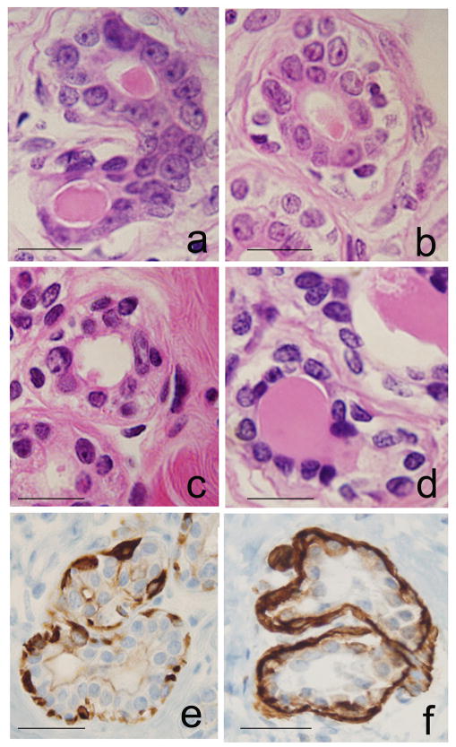
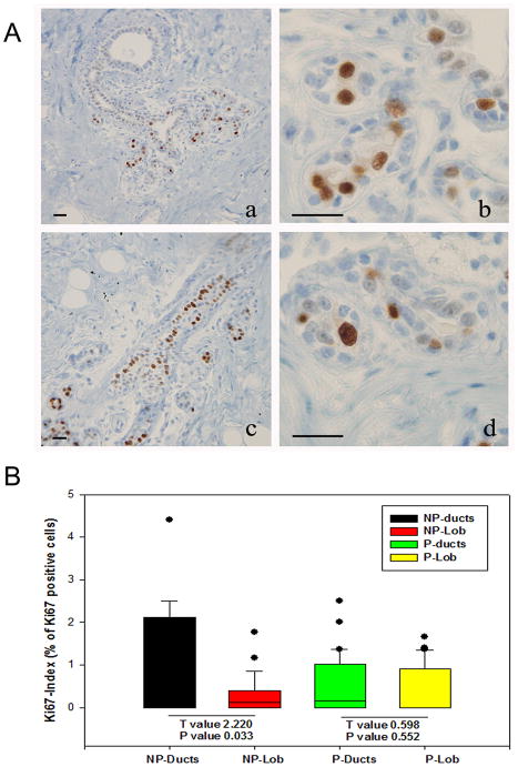
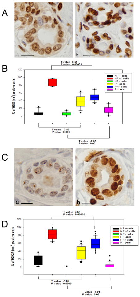
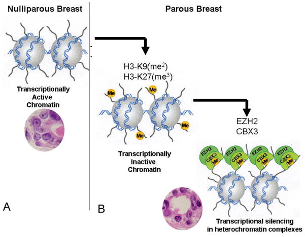
Early pregnancy and multiparity are known to reduce the risk of women to develop breast cancer at menopause. Based on the knowledge that the differentiation of the breast induced by the hormones of pregnancy plays a major role in this protection, this work was performed with the purpose of identifying what differentiation-associated molecular changes persist in the breast until menopause. Core needle biopsies (CNB) obtained from the breast of 42 nulliparous (NP) and 71 parous (P) postmenopausal women were analyzed in morphology, immunocytochemistry and gene expression. Whereas in the NP breast, nuclei of epithelial cells were large and euchromatic, in the P breast they were small and hyperchromatic, showing strong methylation of histone 3 at lysine 9 and 27. Transcriptomic analysis performed using Affymetrix HG_U133 oligonucleotide arrays revealed that in CNB of the P breast, there were 267 upregulated probesets that comprised genes controlling chromatin organization, transcription regulation, splicing machinery, mRNA processing and noncoding elements including XIST. We concluded that the differentiation process induced by pregnancy is centered in chromatin remodeling and in the mRNA processing reactome, both of which emerge as important regulatory pathways. These are indicative of a safeguard step that maintains the fidelity of the transcription process, becoming the ultimate mechanism mediating the protection of the breast conferred by full-term pregnancy.
Copyright © 2011 UICC.
Figures
References
-
- Jemal A, Bray F, Center MM, Ferlay J, Ward E, Forman D. Global Cancer Statistics. CA Cancer J Clin. 2011;61:69–90. - PubMed
-
- Harford JB. Breast-cancer early detection in low-income and middle-income countries: do what you can versus one size fits all. Lancet Oncol. 2011;12:306–12. - PubMed
-
- Hinkula M, Pukkala E, Kyyrönen P, Kauppila A. Grand multiparity and the risk of breast cancer: population-based study in Finland. Cancer Causes Control. 2001;12:491–500. - PubMed
-
- Yang XR, Chang-Claude J, Goode L, Couch FJ, Nevanlinna H, Milne RL, Gaudet M, Schmidt MK, Broeks A, Cox A, Fasching PA, Hein R, et al. Associations of breast cancer risk factors with tumor subtypes: a pooled analysis from the Breast Cancer Association Consortium studies. J Natl Cancer Inst. 2011;103:250–63. - PMC - PubMed
Publication types
MeSH terms
Substances
Grants and funding
LinkOut - more resources
Full Text Sources
Other Literature Sources
Molecular Biology Databases
Miscellaneous
