. 2012;195(1-2):159-70.
doi: 10.1159/000331400. Epub 2011 Oct 24.

Right ventricular outflow tract repair with a cardiac biologic scaffold

Affiliations

Right ventricular outflow tract repair with a cardiac biologic scaffold

John M Wainwright et al. Cells Tissues Organs. 2012.

Abstract

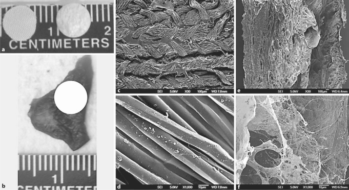
Background: Surgical reconstruction of congenital heart defects is often limited by the nonresorbable material used to approximate normal anatomy. In contrast, biologic scaffold materials composed of resorbable non-cross-linked extracellular matrix (ECM) have been used for tissue reconstruction of multiple organs and are replaced by host tissue. Preparation of whole organ ECM by decellularization through vascular perfusion can maintain much of the native three-dimensional (3D) structure, strength, and tissue-specific composition. A 3D cardiac ECM (C-ECM) biologic scaffold material would logically have structural and functional advantages over materials such as Dacron™ for myocardial repair, but the in vivo remodeling characteristics of C-ECM have not been investigated to date.

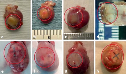
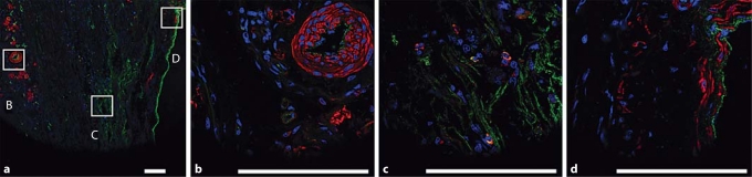
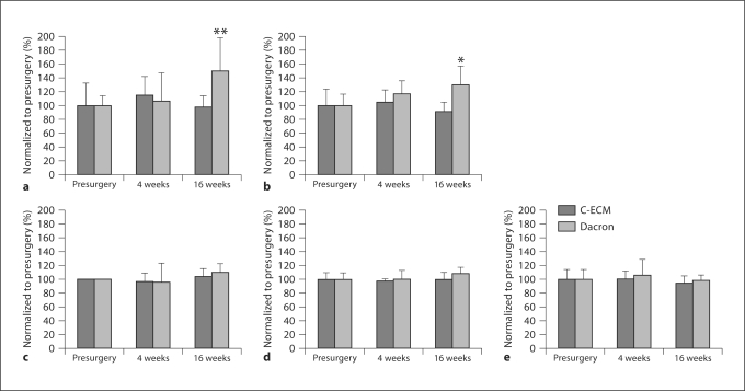
Methods and results: A porcine C-ECM patch or Dacron patch was used to reconstruct a full-thickness right ventricular outflow tract (RVOT) defect in a rat model with end points of structural remodeling function at 16 weeks. The Dacron patch was encapsulated by dense fibrous tissue and showed little cellular infiltration. Echocardiographic analysis showed that the right ventricle of the hearts patched with Dacron were dilated at 16 weeks compared to presurgery baseline values. The C-ECM patch remodeled into dense, cellular connective tissue with scattered small islands of cardiomyocytes. The hearts patched with C-ECM showed no difference in the size or function of the ventricles as compared to baseline values at both 4 and 16 weeks.

Conclusions: The C-ECM patch was associated with better functional and histomorphological outcomes compared to the Dacron patch in this rat model of RVOT reconstruction.


Figures

Fig. 1
Fig. 1
Dacron and C-ECM patches. a Macroscopic photo of the patches. Dacron is on the left and C-ECM is on the right. b Approximate placement of a 6-mm patch in the RVOT shown on excised RV free wall. SEM of Dacron: ×30 (c) and ×1,000 (d). SEM of C-ECM with endocardium on the right side: ×30 (e) and ×1,000 (f).
Fig. 2
Fig. 2
Macroscopic images of the rat heart with the patched area at 2, 4, 8, and 16 weeks postsurgery are enclosed in red ovals. The C-ECM patch decreased in thickness as it remodeled. Fibrous adhesions can be seen at 2 and 4 weeks on the Dacron-patched hearts. The adhesions were removed from the hearts at the later time points so that the patch could be better visualized. C-ECM at 2 (a), 4 (b), 8 (c), and 16 weeks (d) after fixation. Dacron at 2 (e), 4 (f), 8 (g), and 16 weeks (h).
Fig. 3
Fig. 3
Masson's trichrome C-ECM patch at 2, 4, 8, and 16 weeks postsurgery. The C-ECM patch recellularizes and remodels over 16 weeks whereas there are few changes in the Dacron-patched hearts over the 16 weeks. C-ECM at 2 (a), 4 (b), 8 (c), and 16 weeks (d). e C-ECM from the inset in d at 16 weeks. Dacron at 2 (f), 4 (g), 8 (h), and 16 weeks (i). j Dacron from the inset in i at 16 weeks. Scale bar = 500 μm. RV and LV intracavitary spaces are labeled where appropriate.
Fig. 4
Fig. 4
Masson's trichrome and IF staining of the Dacron-patched area at 16 weeks showing little cellular infiltrate of the tissue surrounding the patch and no cellular infiltrate into the Dacron weave. There is an intact endothelium on the endocardial surface but no cardiomyocytes surrounding patch. a ×2, Masson's trichrome. b ×10 magnification with transmission light capture of the inset in a. c ×60 magnification of the inset in b. vWF = Green; α-actinin = red; DRAQ5 = blue. Scale bar = 100 μm.
Fig. 5
Fig. 5
IF staining of the remodeled C-ECM patch showing vWF-positive endothelium and vasculature and SMMHCII-positive smooth muscle throughout the middle of the patch at 16 weeks. a ×10. b ×60 magnification of inset B in a. c ×60 magnification of inset C in a. d ×60 magnification of inset D in a. vWF = Green; SMMHCII = red; DRAQ5 = blue. Scale bar = 100 μm.
Fig. 6
Fig. 6
IF staining of the remodeled C-ECM patch showing α-actinin-positive cardiomyocytes in the middle of the patch on the epicardial surface at 16 weeks. α-Actinin = Red; nuclei = blue. a ×10. b ×40 magnification of the inset in a. Scale bar = 100 μm.
Fig. 7
Fig. 7
IF staining of the remodeled C-ECM patch showing α-actinin and cardiac troponin T-positive cardiomyocytes with connexin 43-labeled gap junctions, vWF-labeled endothelium, and SMMHCII-labeled smooth muscle in the middle of the patched area on the endocardial surface at 16 weeks. a ×10. vWF = Green; α-actinin = red; DAPI = blue. Scale bar = 200 μm. b–e Magnifications of the inset area on sequential slides. Scale bar = 50 μm. b ×63. α-Actinin = Green; cardiac troponin T = red; DRAQ5 = blue. c ×60. vWF = Green; α-actinin = red; DAPI = blue. d ×63. Connexin 43 = Green; α-actinin = red; DRAQ5 = blue. e ×60. SMMHCII = Green; α-actinin = red; DAPI = blue.
Fig. 8
Fig. 8
Echocardiographic analysis at presurgery and 4 and 16 weeks postsurgery with one standard deviation. a RV minimum diameter. b RV maximum diameter. c LV minimum diameter. d LV maximum diameter. e LV fractional area shortening diameter. For the C-ECM patch, none of the measures were different from presurgery values at either 4 or 16 weeks. For the Dacron patch the RV minimum and maximum diameters had dilated by 16 weeks. * p < 0.05 compared to presurgery; ** p < 0.05 compared to presurgery and 4 weeks postsurgery.

References

    1. Badylak S.F., Kochupura P.V., Cohen I.S., Doronin S.V., Saltman A.E., Gilbert T.W., Kelly D.J., Ignotz R.A., Gaudette G.R. The use of extracellular matrix as an inductive scaffold for the partial replacement of functional myocardium. Cell Transplant. 2006;15(suppl 1):S29–S40. - PubMed
    1. Badylak S.F., Kokini K., Tullius B., Simmons-Byrd A., Morff R. Morphologic study of small intestinal submucosa as a body wall repair device. J Surg Res. 2002;103:190–202. - PubMed
    1. Badylak S.F., Liang A., Record R., Tullius R., Hodde J.P. Endothelial cell adherence to small intestinal submucosa: an acellular bioscaffold. Biomaterials. 1999;20:2257–2263. - PubMed
    1. Badylak S.F., Park K., Peppas N., McCabe G., Yoder M. Marrow-derived cells populate scaffolds composed of xenogeneic extracellular matrix. Exp Hematol. 2001;29:1310–1318. - PubMed
    1. Badylak S.F., Vorp D.A., Spievack A.R., Simmons-Byrd A., Hanke J., Freytes D.O., Thapa A., Gilbert T.W., Nieponice A. Esophageal reconstruction with ECM and muscle tissue in a dog model. J Surg Res. 2005;128:87–97. - PubMed

Publication types

Substances