Comparative Study
. 2011 Dec 1;29(34):4482-90.
doi: 10.1200/JCO.2010.33.8715. Epub 2011 Oct 24.

Evidence for sequenced molecular evolution of IDH1 mutant glioblastoma from a distinct cell of origin

Affiliations
Comparative Study

Evidence for sequenced molecular evolution of IDH1 mutant glioblastoma from a distinct cell of origin

Albert Lai et al. J Clin Oncol. .

Abstract

Purpose: Mutation in isocitrate dehydrogenase 1 (IDH1) at R132 (IDH1(R132MUT)) is frequent in low-grade diffuse gliomas and, within glioblastoma (GBM), has been proposed as a marker for GBMs that arise by transformation from lower-grade gliomas, regardless of clinical history. To determine how GBMs arising with IDH1(R132MUT) differ from other GBMs, we undertook a comprehensive comparison of patients presenting clinically with primary GBM as a function of IDH1(R132) mutation status.

Patients and methods: In all, 618 treatment-naive primary GBMs and 235 lower-grade diffuse gliomas were sequenced for IDH1(R132) and analyzed for demographic, radiographic, anatomic, histologic, genomic, epigenetic, and transcriptional characteristics.

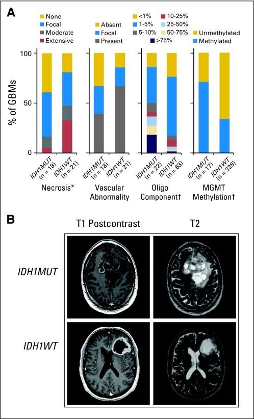
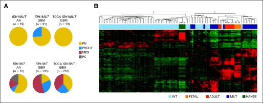
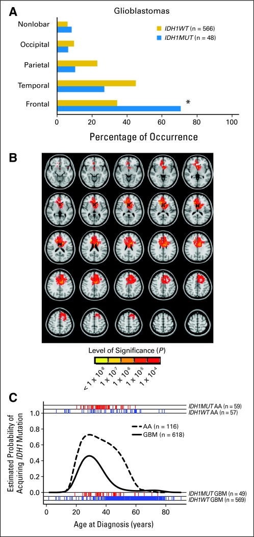
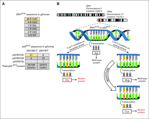
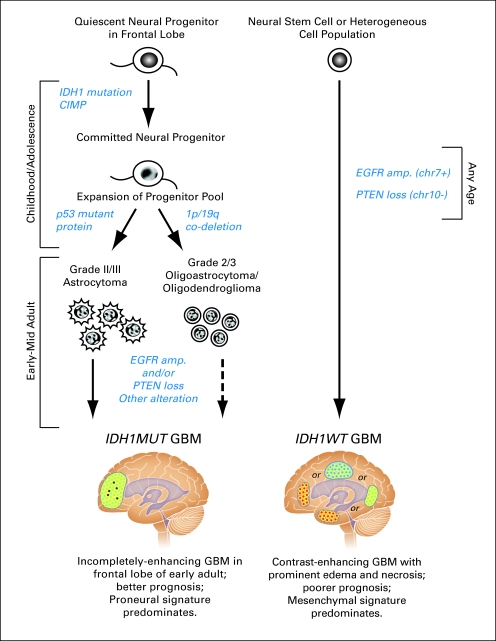
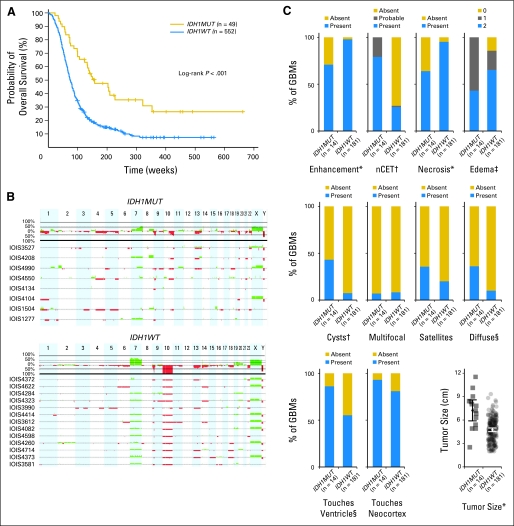
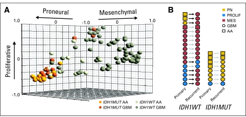
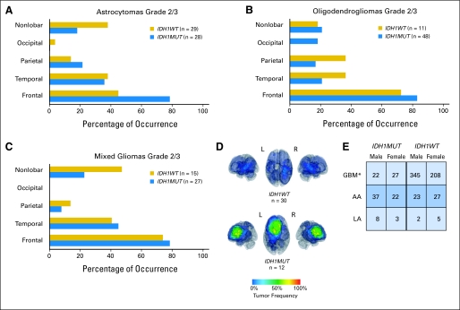
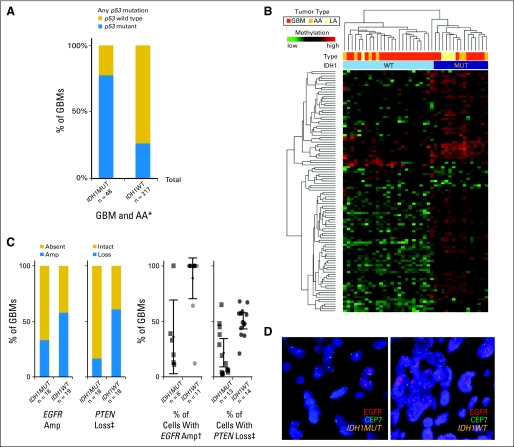
Results: Investigation revealed a constellation of features that distinguishes IDH1(R132MUT) GBMs from other GBMs (including frontal location and lesser extent of contrast enhancement and necrosis), relates them to lower-grade IDH1(R132MUT) gliomas, and supports the concept that IDH1(R132MUT) gliomas arise from a neural precursor population that is spatially and temporally restricted in the brain. The observed patterns of DNA sequence, methylation, and copy number alterations support a model of ordered molecular evolution of IDH1(R132MUT) GBM in which the appearance of mutant IDH1 protein is an initial event, followed by production of p53 mutant protein, and finally by copy number alterations of PTEN and EGFR.

Conclusion: Although histologically similar, GBMs arising with and without IDH1(R132MUT) appear to represent distinct disease entities that arise from separate cell types of origin as the result of largely nonoverlapping sets of molecular events. Optimal clinical management should account for the distinction between these GBM disease subtypes.


Conflict of interest statement

Authors' disclosures of potential conflicts of interest and author contributions are found at the end of this article.

Figures

Fig 1.
Fig 1.
Glioblastomas (GBMs) with mutations in isocitrate dehydrogenase 1 (IDH1) at R132 (IDH1R132MUT; IDH1MUT) are phenotypically distinct from IDH1R132-wild type (IDH1R132WT; IDH1WT) GBMs. (A) Histologic assessment of IDH1R132MUT and IDH1R132WT GBMs reveals decreased necrosis (*P < .05) and oligodendroglial content (†P < .005), with a trend toward decreased vascular abnormalities (P < .075) for IDH1R132MUT GBMs (Data Supplement). Frequency of O6-methylguanine–DNA methyltransferase (MGMT) promoter methylation is increased in IDH1R132MUT GBMs (†P < .005; Data Supplement). (B) Representative pretreatment contrast-enhanced T1-weighted images (left) with corresponding T2-weighted images (right) of a patient with IDH1R132MUT (IDH1MUT; upper panel) and a patient with IDH1R132WT (IDH1WT; lower panel).
Fig 2.
Fig 2.
Glioblastomas (GBMs) with mutations in isocitrate dehydrogenase 1 (IDH1) at R132 (IDH1R132MUT; IDH1MUT) are restricted from Mesenchymal gene expression and show greater similarity to transcriptional signatures of brain cells than to those of neural stem cells. (A) Gene expression analysis reveals a robust positive association of IDH1R132MUT high-grade astrocytomas with the Proneural signature and an absolute exclusion of the Mesenchymal gene signature from IDH1R132MUT tumors. Subtypes are labeled as follows: Proneural (PN), Proliferative (PROLIF), and Mesenchymal (MES). PC denotes poorly classified tumors. Pie charts depict tabulated data for all tumors assigned to subtype. AA, anaplastic astrocytoma (grade 3 astrocytoma) cohort G (Data Supplement); GBM, cohorts A-D, and F (Data Supplement); TCGA, newly diagnosed GBM samples from The Cancer Genome Atlas (TCGA) data set (Cancer Genome Atlas Research Network: Nature 455:1061-1068, 2008). (B) Heatmap of hierarchical clustering of GBMs in cohort C based on gene list derived from most variable genes between IDH1R132MUT and IDH1R132-wild type (IDH1R132WT; IDH1WT) GBMs (Data Supplement). IDH1R132WT GBMs (WT) show similarity to human adult neural stem cells for experimentation (HANSE), whereas IDH1R132MUT GBMs (MUT) show similarity to fetal/adult brain tissue (FETAL, ADULT).
Fig 3.
Fig 3.
Spatial and temporal restriction of gliomas arising with mutations in isocitrate dehydrogenase 1 (IDH1) at R132 (IDH1R132MUT; IDH1MUT). (A) IDH1R132MUT de novo glioblastomas (GBMs) occur more frequently in the frontal lobe compared with IDH1R132-wild type (IDH1R132WT; IDH1WT) GBMs (*P < .001, two-tailed Fisher's exact test, with Bonferroni correction for multiple comparisons; Data Supplement). (B) Area of differential involvement (ADIFFI) analysis of axial magnetic resonance images. Voxels that are more frequently involved in IDH1R132MUT GBMs can be seen to cluster in the frontal lobe near the rostral extension of the lateral ventricle. All voxels with P < .0001 for differential involvement are depicted, as indicated in the color-coded bar. (C). IDH1R132MUT GBMs are temporally restricted. Logistic regression curves show the probability of IDH1R132MUT occuring in anaplastic astrocytomas (AAs; grade 3 astrocytoma) and GBMs as a function of age. The red (IDH1R132MUT) and blue (IDH1R132WT) tick marks show actual ages of patients (Data Supplement).
Fig 4.
Fig 4.
Evidence for ordered appearance of aberrations in isocitrate dehydrogenase 1 (IDH1) mutant glioblastoma (GBM). (A) Top: Frequencies of sequences found at IDH1R132 show that the predominant substitution is His (yellow box). Bottom: Among gliomas with mutations at IDH1R132 (IDH1MUT) and p53R273, a predominance of Cys substitution at p53R273 is seen (yellow box); for IDH1R132-wild type (IDH1WT) tumors, the preferred substitution at p53R273 is His. (B) Schematic diagram showing that IDH1 mutation on the template strand and p53 mutation on coding strand can select for the expression of IDH1 mutant protein before p53 mutant protein if replication is delayed.
Fig 5.
Fig 5.
Model comparing glioblastomas (GBMs) arising with and without mutations in isocitrate dehydrogenase 1 (IDH1) at R132. We propose that the IDH1R132MUT GBM pathway (left) is initiated by the occurrence of IDH1 mutation and resultant CpG island methylator phenotype (CIMP) in a quiescent neural progenitor residing in the frontal lobe. Although p53 mutation can be present during this time, expression of p53 mutant protein ensues only after expansion of this progenitor pool during late adolescence or early adulthood. According to the proposed model, glioma formation along the IDH1R132MUT pathway requires the ordered appearance of IDH1 mutant protein and CIMP, followed by p53 mutant protein (or 1p/19q co-deletion). Tumors along this pathway arise from a spatially and temporally restricted neural progenitor population and most frequently maintain a Proneural gene expression signature. Transformation to IDH1R132MUT GBM requires EGFR amplification (amp), PTEN deletion, or other genomic alterations. In contrast, in the IDH1R132WT GBM pathway (right), EGFR amplification, and PTEN loss frequently act in concert to drive GBM formation from a cell population that maintains the ability to adopt a Mesenchymal gene expression signature. chr7+, chromosome 7 gain; chr10−, chromosome 10 loss. IDH1MUT, IDH1R132MUT; IDH1WT, IDH1R132WT.
Fig A1.
Fig A1.
Glioblastomas (GBMs) with mutations in isocitrate dehydrogenase 1 (IDH1) at R132 (IDH1R132MUT; IDH1MUT) are phenotypically distinct from IDH1R132-wild type (IDH1R132WT; IDH1WT) GBMs. (A) Kaplan-Meier survival curves of adult patients with de novo GBMs depict increased overall survival in patients with IDH1R132MUT versus IDH1R132WT tumors (log-rank test P < .001). Tick marks denote censored patients. All newly diagnosed adult (≥ 18 years) patients with available survival data are included in this analysis (Data Supplement). (B) Relative DNA copy number estimates of a sampling of IDH1R132MUT GBMs (upper panel) and IDH1R132WT GBMs (lower panel) demonstrates a minority of instances of co-deletion of 1p/19q in IDH1R132H GBMs (Data Supplement). Green and red deflections from the x axis indicate copy number gains and losses, respectively. The top portion of each panel depicts the percentage of tumors surveyed that exhibited copy number change events. The bottom traces in each panel show, for each sample (identified by case ID), a ratio of the fold change in intensity signals between the test and reference sample, transformed by using log2. (C) Scoring of a panel of radiologic variables in a sampling of GBMs reveals decreased occurrence of enhancement (*P < .005), increased frequency of non–contrast-enhancing tumor (nCET; †P < .001), decreased incidence of necrosis (*P < .005), decreased extent of edema (‡P < .01), increased presence of cysts (†P < .001), increased incidence of diffuse component (§P < .05), increased frequency of touches ventricle (§P < .05), and increased tumor size (*P < .005) for IDH1R132MUT compared with IDH1R132WT; Data Supplement). Occurrences of multifocal lesions, satellites, and touches neocortex were not different.
Fig A2.
Fig A2.
Glioblastomas (GBMs) with mutations in isocitrate dehydrogenase 1 (IDH1) at R132 (IDH1R132MUT; IDH1MUT) are restricted from Mesenchymal gene expression. Gene expression analysis reveals a robust positive association of IDH1R132MUT high-grade astrocytomas with the Proneural signature and an absolute exclusion of the Mesenchymal gene signature from IDH1R132MUT tumors. Subtypes and their respective centroids are labeled as follows: Proneural (PN), Proliferative (PROLIF), and Mesenchymal (MES). (A) The three-dimensional graphical plot illustrates the similarity between individual tumor samples in cohorts C and G to each of the three centroids created by K means clustering to define tumor subtypes. (B) Matched primary and recurrent pairs of tissue show that IDH1R132MUT GBMs and anaplastic astrocytomas (AAs; grade 3 astrocytoma) fail to acquire a Mesenchymal gene expression signature on recurrence (Data Supplement). IDH1WT, IDH1R132WT.
Fig A3.
Fig A3.
Spatial restriction of gliomas arising with mutations in isocitrate dehydrogenase 1 (IDH1) at R132 (IDH1R132MUT; IDH1MUT). Location of grade 2 and 3 astrocytomas (A), oligodendrogliomas (B), and mixed gliomas (C) show that, as for GBMs, the majority of IDH1R132MUT gliomas of these histologic subtypes involve the frontal lobe (Data Supplement). (D) Three-dimensional representations of frequency maps generated by overlaying tumor regions determined from digital magnetic resonance images of patient tissue with IDH1R132-wild type (IDH1R132WT; IDH1WT) and IDH1R132MUT GBMs, depicting frequent frontal lobe involvement in patients with IDH1R132MUT tumors. In contrast to IDH1R132MUT GBMs, IDH1R132WT GBMs are more broadly distributed throughout the forebrain (Data supplement). (E) Gender distribution of adult patients with astrocytoma as a function of tumor grade and IDH1R132 mutation status. *P < .05 for IDH1R132MUT versus IDH1R132WT GBM. AA, anaplastic astrocytoma; LA, low-grade astrocytoma.
Fig A4.
Fig A4.
Evidence for ordered appearance of aberrations in isocitrate dehydrogenase 1 (IDH1) mutant glioblastoma (GBM). (A) Tumors with a mutation in IDH1 at R132 (IDH1R132MUT; IDH1MUT) show increased mutation frequency of p53 at any site within exons 4 to 10 in a series of GBMs and anaplastic astrocytomas (AAs; grade 3 astrocytoma) compared with IDH1R132-wild type (IDH1R132WT; IDH1WT) tumors (*P < .001; Data Supplement). (B) Unsupervised hierarchical clustering of DNA methylation data profiling of a series of glioma samples reveals the presence of a CpG island methylator phenotype strongly correlated with IDH1R132MUT (Data Supplement). (C) Quantitative evaluation of fluorescent in situ hybridization for EGFR and PTEN in a series of newly diagnosed GBMs (Data Supplement). Bar graphs (left) depict a nonsignificant trend toward less frequent EGFR amplification (Amp; P < .191) and reduced frequency of cases with IDH1R132MUT GBM showing PTEN loss (+P < .05). Scatter plots (right) show the frequency of cells within individual cases that show these copy number alterations, indicating decreased percentage of cells with EGFR amplification (†P < .001) and PTEN loss (‡P < .05) in IDH1R132MUT GBMs. Cases in which no cells showed detectable alterations are not included in the scatter plots or statistical analysis of percentage of cells. (D) Representative example of fluorescent in situ hybridization using probes to EGFR (red) and to the centromere of chromosome 7 (CEP7, green), showing an example of focal amplification in an IDH1R132MUT GBM and widespread amplification in an IDH1R132WT GBM. LA, low-grade astrocytoma; MUT, mutant; WT, wild type.

Comment in

References

    1. Clarke J, Butowski N, Chang S. Recent advances in therapy for glioblastoma. Arch Neurol. 2010;67:279–283. - PubMed
    1. Hambardzumyan D, Squatrito M, Carbajal E, et al. Glioma formation, cancer stem cells, and akt signaling. Stem Cell Rev. 2008;4:203–210. - PubMed
    1. Alcantara Llaguno S, Chen J, Kwon CH, et al. Malignant astrocytomas originate from neural stem/progenitor cells in a somatic tumor suppressor mouse model. Cancer Cell. 2009;15:45–56. - PMC - PubMed
    1. Alcantara Llaguno SR, Chen J, Parada LF. Signaling in malignant astrocytomas: Role of neural stem cells and its therapeutic implications. Clin Cancer Res. 2009;15:7124–7129. - PMC - PubMed
    1. Ohgaki H, Dessen P, Jourde B, et al. Genetic pathways to glioblastoma: A population-based study. Cancer Res. 2004;64:6892–6899. - PubMed

Publication types

Substances