. 2011 Nov 21;208(12):2393-401.
doi: 10.1084/jem.20110525. Epub 2011 Oct 24.

Dissociation of VE-PTP from VE-cadherin is required for leukocyte extravasation and for VEGF-induced vascular permeability in vivo

Affiliations

Dissociation of VE-PTP from VE-cadherin is required for leukocyte extravasation and for VEGF-induced vascular permeability in vivo

Andre Broermann et al. J Exp Med. .

Abstract

We have recently shown that vascular endothelial protein tyrosine phosphatase (VE-PTP), an endothelial membrane protein, associates with VE-cadherin and is required for optimal VE-cadherin function and endothelial cell contact integrity. The dissociation of VE-PTP from VE-cadherin is triggered by vascular endothelial growth factor (VEGF) and by the binding of leukocytes to endothelial cells in vitro, suggesting that this dissociation is a prerequisite for the destabilization of endothelial cell contacts. Here, we show that VE-cadherin/VE-PTP dissociation also occurs in vivo in response to LPS stimulation of the lung or systemic VEGF stimulation. To show that this dissociation is indeed necessary in vivo for leukocyte extravasation and VEGF-induced vascular permeability, we generated knock-in mice expressing the fusion proteins VE-cadherin-FK 506 binding protein and VE-PTP-FRB* under the control of the endogenous VE-cadherin promoter, thus replacing endogenous VE-cadherin. The additional domains in both fusion proteins allow the heterodimeric complex to be stabilized by a chemical compound (rapalog). We found that intravenous application of the rapalog strongly inhibited VEGF-induced (skin) and LPS-induced (lung) vascular permeability and inhibited neutrophil extravasation in the IL-1β inflamed cremaster and the LPS-inflamed lung. We conclude that the dissociation of VE-PTP from VE-cadherin is indeed required in vivo for the opening of endothelial cell contacts during induction of vascular permeability and leukocyte extravasation.


Figures
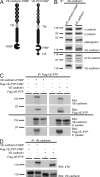
Figure 1.
Figure 1.
VE-cadherin-FKPB and Flag-VE-PTP-FRB* show the same characteristics as WT proteins. (A) Schematic illustration of VE-cadherin-FKBP and VE-PTP-FRB* transmembrane domain (TM) and HA-tag (HA). (B) VE-cadherin or VE-cadherin-FKBP were expressed in COS-7 cells. VE-cadherin was immunoprecipitated, and the indicated proteins were detected by immunoblotting. The horizontal line indicates different experiments. (C) The indicated proteins were expressed in COS-7 cells, and Flag-VE-PTP or Flag-VE-PTP-FRB* were immunoprecipitated, followed by analysis for Flag-VE-PTP and co-precipitated VE-cadherin-FKBP or VE-cadherin by immunoblotting. Expression of transfected proteins was controlled by immunoblots of total cell lysates. (D) VEGFR-2 was coexpressed with the indicated proteins in COS-7 cells. VE-cadherin was immunoprecipitated, followed by immunoblotting for phosphotyrosine and VE-cadherin. Data are representative for two (B), six (C), and three (D) independent experiments.
Figure 2.
Figure 2.
Generation of transgenic mice expressing VE-cadherin-FKBP and VE-PTP-FRB*. (A) Schematic illustration of the RMCE approach. The gene replacement cassette contains cDNAs for VE-cadherin-FKBP and full-length VE-PTP-FRB* separated by an IRES site, as well as a Hygromycin cassette, flanked by two FRT sites. The cassette is flanked by two incompatible loxP sites. Using RMCE, the ATG-containing exon 2 of VE-cadherin was replaced by the cDNA insertion cassette. Positions of the primers for PCR-genotyping are indicated. (B) Lung lysates of either wild-type (+/+), heterozygous (+/KI), or homozygous mutant (KI/KI) mice were immunoblotted for VE-cadherin and as loading control for Tie-2. (C) Endogenous VE-PTP was immunoprecipitated with anti–VE-PTP-C and VE-PTP-FRB* was immunoprecipitated with anti-HA antibodies from lung lysates of homozygous KI (KI/KI) or WT (+/+) mice. Immunoprecipitations with goat IgG (CoIgG) were performed as a control. Precipitates were analyzed by immunoblotting with antibodies against the extracellular domain of VE-PTP. Note that KI mice showed only slightly increased overall amounts of VE-PTP compared with WT mice (left) because the anti–VE-PTP-C antibodies bind to VE-PTP-FRB* only very weakly. (D) Whole mount staining of venules in cremaster from WT mice (left) and KI mice (right) for VE-cadherin or VE-cadherin-FKBP, respectively. Bars, 20 µm. Data are representative for three (B and C) and two (D) independent experiments.
Figure 3.
Figure 3.
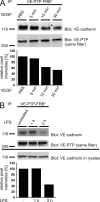
Dissociation of VE-PTP FRB* from VE-cadherin-FKBP occurs in vivo after application of VEGF or LPS. (A) Homozygous KI mice received 3 µg VEGF i.v. and were sacrificed at the indicated time points. VE-PTP FRB* was immunoprecipitated from lung lysates and precipitates were blotted for VE-PTP and VE-cadherin. (B) KI mice were untreated or inhaled an LPS aerosol for 1 h (3.5 mg/7 ml). LPS-treated mice were sacrificed either immediately after LPS exposure or left untreated for an additional 1 h. Lungs were analyzed at the indicated time points after starting LPS treatment. Degradation of VE-cadherin was controlled by immunoblotting total lung lysates (bottom). Data are representative of three independent experiments (A and B).
Figure 4.
Figure 4.
Dissociation of VE-PTP from VE-cadherin is necessary for VEGF-induced permeability in vivo. (A) Homozygous KI mice were injected i.v. either with vehicle alone (left) or with rapalog (right) 8 and 4 h before the assay. Mice were then i.v. injected with either PBS or with VEGF (as indicated), and 30 min later lung lysates were immunoprecipitated for VE-PTP-FRB*. Precipitated material was analyzed by immunoblotting for VE-cadherin-FKBP and VE-PTP expression. To control for degradation, aliquots of total lung lysates were set aside and analyzed by immunoblotting for VE-cadherin-FKBP (bottom). The amount of immunoprecipitated VE-PTP-FRB and co-precipitated VE-cadherin-FKBP detected from VEGF-treated mice is shown as percentage of the amount precipitated from PBS-treated mice (numbers below). Graphs in inserts give co-precipitation efficiency. Note that the rapalog blocked dissociation of VE-cadherin from VE-PTP. (B) Homozygous KI mice were treated with rapalog or vehicle and subsequently with VEGF as in A. Lung lysates were immunoprecipitated for VE-cadherin and precipitates were immunoblotted first for phosphotyrosine, and then for VE-cadherin and plakoglobin (γ-catenin). (C) Lung lysates from KI mice were immunoprecipitated for Tie-2 and immunoblots were analyzed for phospho-tyrosine and subsequently for Tie-2. (D) Homozygous KI mice were injected i.v. with rapalog (black bars) or vehicle alone (white bars) 8 and 4 h before the assay. At the start of the assay, Evan’s blue was i.v. injected, followed by intradermal injection of VEGF or PBS 10 min later. After 30 min, mice were sacrificed and the dye was extracted from skin samples and quantified. n = 5 mice per group. Data are representative of two (A–C) or three (D) independent experiments. (E) Homozygous KI mice were treated with rapalog or vehicle as in D, followed by exposing the mice to nebulized LPS for 45 min. 4 h later, lungs were lavaged and protein content was measured. n = 3 mice per group. Results shown are representative of two independent experiments. *, P ≤ 0.05; **, P ≤ 0.01.
Figure 5.
Figure 5.
Dissociation of VE-PTP from VE-cadherin is necessary for efficient transendothelial migration of leukocytes in vivo. Homozygous KI mice were i.v. injected with either rapalog or vehicle alone 8 and 4 h before the assay, followed by an intrascrotal injection of 50 ng IL-1β. The cremaster muscle was prepared 4 h later for intravital imaging. Numbers of extravasated (A) and adherent leukocytes (B), rolling flux fraction (C), and rolling velocity (D) were determined. The results are displayed as mean ± SEM of at least 30 vessels from five independent animals in each group. (E) Homozygous KI mice were treated with rapalog or vehicle and exposed to nebulized LPS as in Fig. 4 E. The leukocytes in the bronchoalveolar lavage fluid were counted and the numbers of neutrophils were determined by FACS. n = 3 mice per group. Results shown are representative for two independent experiments. *, P ≤ 0.05; **, P ≤ 0.01; ***, P ≤ 0.001.

References

    1. Allingham M.J., van Buul J.D., Burridge K. 2007. ICAM-1-mediated, Src- and Pyk2-dependent vascular endothelial cadherin tyrosine phosphorylation is required for leukocyte transendothelial migration. J. Immunol. 179:4053–4064 - PubMed
    1. Bäumer S., Keller L., Holtmann A., Funke R., August B., Gamp A., Wolburg H., Wolburg-Buchholz K., Deutsch U., Vestweber D. 2006. Vascular endothelial cell-specific phosphotyrosine phosphatase (VE-PTP) activity is required for blood vessel development. Blood. 107:4754–4762 10.1182/blood-2006-01-0141 - DOI - PubMed
    1. Bixel G., Kloep S., Butz S., Petri B., Engelhardt B., Vestweber D. 2004. Mouse CD99 participates in T-cell recruitment into inflamed skin. Blood. 104:3205–3213 10.1182/blood-2004-03-1184 - DOI - PubMed
    1. Breviario F., Caveda L., Corada M., Martin-Padura I., Navarro P., Golay J., Introna M., Gulino D., Lampugnani M.G., Dejana E. 1995. Functional properties of human vascular endothelial cadherin (7B4/cadherin-5), an endothelium-specific cadherin. Arterioscler. Thromb. Vasc. Biol. 15:1229–1239 10.1161/01.ATV.15.8.1229 - DOI - PubMed
    1. Carman C.V., Sage P.T., Sciuto T.E., de la Fuente M.A., Geha R.S., Ochs H.D., Dvorak H.F., Dvorak A.M., Springer T.A. 2007. Transcellular diapedesis is initiated by invasive podosomes. Immunity. 26:784–797 10.1016/j.immuni.2007.04.015 - DOI - PMC - PubMed

Publication types

MeSH terms