

Gold nanocages as contrast agents for photoacoustic imaging
- PMID: 22025337
- PMCID: PMC6942690
- DOI: 10.1002/cmmi.439
Gold nanocages as contrast agents for photoacoustic imaging
Abstract
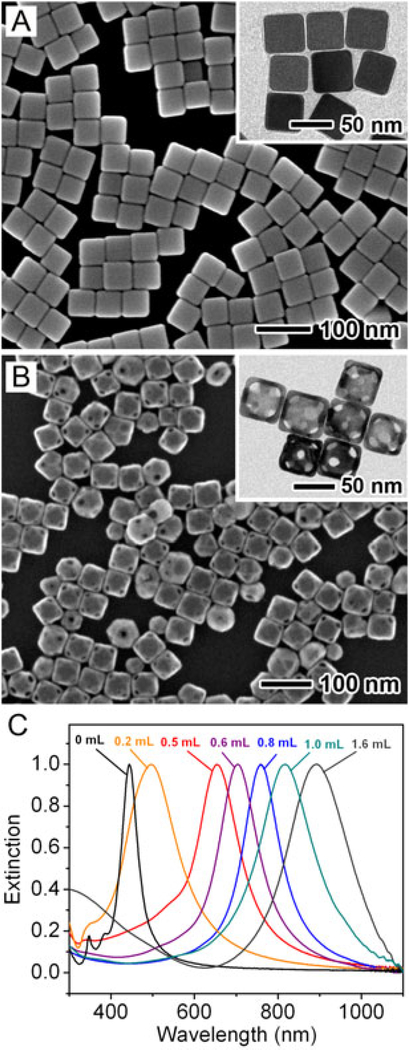
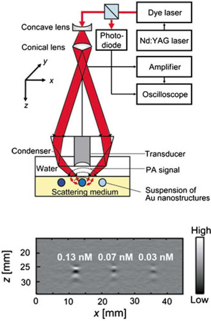
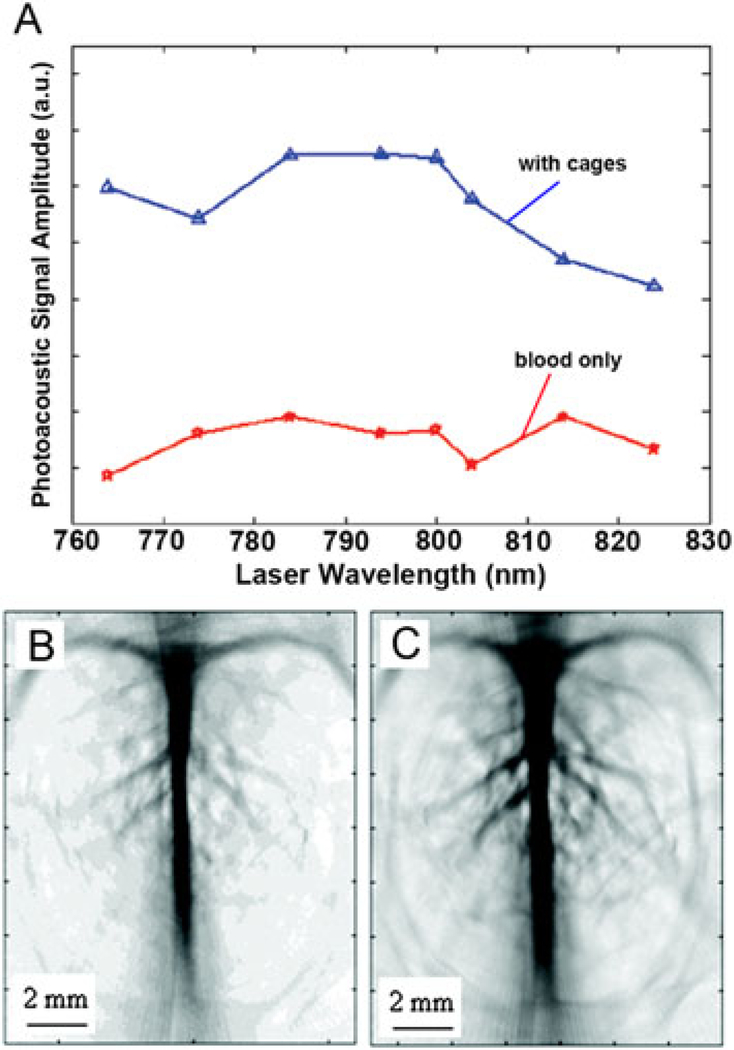
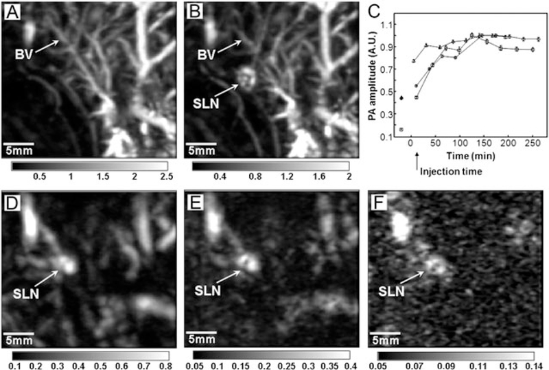
Gold nanoparticles with tunable absorption and scattering properties have been developed as contrast agents for various optical imaging techniques. As a hybrid modality that combines the merits of both optical and ultrasonic imaging, photoacoustic (PA) imaging also benefits from the use of these nanoparticles to greatly enhance the contrast for visualization of structures and biomarkers in biological tissues. Gold nanocages characterized by hollow interiors, ultrathin and porous walls are of particular interest for in vivo PA imaging because of their compact sizes, bio-inertness and well-defined surface chemistry, as well as their strong and highly wavelength-tunable optical absorption in the near-infrared (NIR) optical window of soft tissues. This review discusses the application of gold nanocages as a new class of contrast agents for PA imaging in the context of cancer diagnosis.
Copyright © 2011 John Wiley & Sons, Ltd.
Figures

References
-
- Wang LV, Wu H. Biomedical Optics: Principles and Imaging, John Wiley and Sons, Hoboken, NJ, 2007.
-
- Grinvald A, Lieke E, Frostig RD, Gilbert CD, Wiesel TN. Functional architecture of cortex revealed by optical imaging of intrinsic signals. Nature 1986; 324: 361–364. - PubMed
-
- Rapacholi MH. Essentials of Medical Ultrasound: A Practical Introduction to the Principles, Techniques and Biomedical Applications. Humana: New York, 1982.
-
- Wang XD, Pang YJ, Ku G, Xie XY, Stoica G, Wang LV. Noninvasive laser- induced photoacoustic tomography for structural and functional in vivo imaging of the brain. Nat Biotechnol 2003; 21: 803–806. - PubMed
Publication types
MeSH terms
Substances
Grants and funding
LinkOut - more resources
Full Text Sources
Medical
Miscellaneous
