. 2011 Nov 29;124(22):2411-22.
doi: 10.1161/CIRCULATIONAHA.111.040071. Epub 2011 Oct 24.

Distinct epigenomic features in end-stage failing human hearts

Affiliations

Distinct epigenomic features in end-stage failing human hearts

Mehregan Movassagh et al. Circulation. .

Abstract

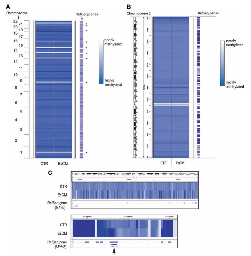
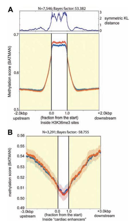
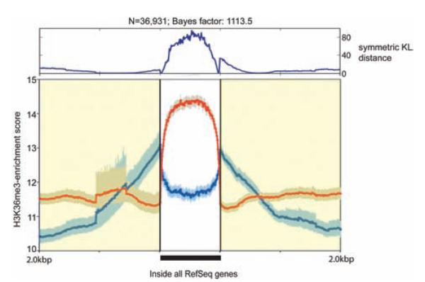
Background: The epigenome refers to marks on the genome, including DNA methylation and histone modifications, that regulate the expression of underlying genes. A consistent profile of gene expression changes in end-stage cardiomyopathy led us to hypothesize that distinct global patterns of the epigenome may also exist.

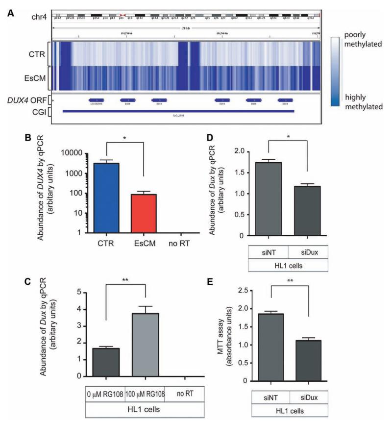
Methods and results: We constructed genome-wide maps of DNA methylation and histone-3 lysine-36 trimethylation (H3K36me3) enrichment for cardiomyopathic and normal human hearts. More than 506 Mb sequences per library were generated by high-throughput sequencing, allowing us to assign methylation scores to ≈28 million CG dinucleotides in the human genome. DNA methylation was significantly different in promoter CpG islands, intragenic CpG islands, gene bodies, and H3K36me3-enriched regions of the genome. DNA methylation differences were present in promoters of upregulated genes but not downregulated genes. H3K36me3 enrichment itself was also significantly different in coding regions of the genome. Specifically, abundance of RNA transcripts encoded by the DUX4 locus correlated to differential DNA methylation and H3K36me3 enrichment. In vitro, Dux gene expression was responsive to a specific inhibitor of DNA methyltransferase, and Dux siRNA knockdown led to reduced cell viability.

Conclusions: Distinct epigenomic patterns exist in important DNA elements of the cardiac genome in human end-stage cardiomyopathy. The epigenome may control the expression of local or distal genes with critical functions in myocardial stress response. If epigenomic patterns track with disease progression, assays for the epigenome may be useful for assessing prognosis in heart failure. Further studies are needed to determine whether and how the epigenome contributes to the development of cardiomyopathy.


Figures

Figure 1
Figure 1
DNA methylation profiles between control (CTR) and end-stage cardiomyopathic (EsCM) hearts are similar at low resolution (A and B), but distinct differences are found at the level of individual genes (C) and at specific CpG islands (An example is shown with the arrow in C). Averaged DNA methylation profiles were generated for control and EsCM from 8 sequencing libraries comprising 4 normal and 4 EsCM hearts and scored at 100-bp resolution genome wide. Analysis was technically not possible in some gene-poor regions where a high density of repetitive elements was present (asterisk in A).
Figure 2
Figure 2
DNA methylation density plots for all CpG islands (CGIs; A) and subclasses of CGIs (B through E). A, DNA methylation scores (BATMAN) for 27 639 CGIs and CGI shores (3 kb upstream and downstream of CGIs) were plotted for control (CTR; blue) and end-stage cardiomyopathic (EsCM; red; light blue and light red error bars represent bayesian credible intervals for CTR and EsCM, respectively). A Gaussian process 2-sample test returned a log Bayes factor of 15.9 (A positive log Bayes factor implies that the difference between control and EsCM was statistically significant). The symmetrical Kulback-Lieber (KL) divergence was calculated for each position across the x axis and charted above the methylation density plots as symmetrical KL distance and indicates that the methylation profiles differed both at the center of CGIs and at CGI shores. Significant differences in methylation profiles were also found in (B) promoter CGIs and (C) intragenic CGIs but not in (D) intergenic CGIs and (E) 3′ untranslated region (UTR) CGIs. However in the first 2 subclasses, methylation profiles were significantly different at CGIs but not CGI shores.
Figure 3
Figure 3
DNA methylation was significantly different in promoters of (A) upregulated genes but not (B) downregulated genes.
Figure 4
Figure 4
DNA methylation was significantly different in (A) actively transcribed regions of the cardiac genome but not (B) cardiac enhancers. Regions of active transcription in the cardiac genome were determined by H3K36me3 chromatin immunoprecipitation sequencing (ChIP-seq), and regions of cardiac enhancers were obtained from a previous p300 ChIP-seq.
Figure 5
Figure 5
H3K36me3 enrichment profiles within regions of annotated Reference Sequence (RefSeq) genes were significantly different between control (CTR) and end-stage cardiomyopathy (EsCM). Composite H3K36me3 enrichment profiles were generated for control (blue) and EsCM (red) from 2 pooled sequencing libraries comprising 4 normal and 3 EsCM hearts, respectively (see Table I in the online-only Data Supplement for details of samples). Enrichment scores were determined as tag counts per 500 bp per 1 million reads for each sequencing library and analyzed for all 36 109 RefSeq genes (as annotated on the UCSC Genome Browser) and 3 kb upstream and downstream of these regions.
Figure 6
Figure 6
Differential DNA methylation and expression of DUX4 in human hearts and Dux in the mouse HL1 cardiac cell line. A, The intronless DUX4 open reading frame is embedded within the subtelomeric array of D4Z4 repeat units. The CpG island (CGI) in this locus is hypermethylated in end-stage cardiomyopathy (EsCM) hearts but not in control (CTR). B, The abundance of DUX4 RNA transcripts was quantified with DNase-treated RNA from a panel of left ventricular tissue (8 control and 16 EsCM; Table I in the online-only Data Supplement) and normalized by geNorm that was generated with 2 housekeeping genes, RPLPO and TBP. Absent polymerase chain reaction (PCR) products in “no reverse transcriptase (RT) ” controls excluded the likelihood of genomic DNA amplification. *P<0.05. C, The mouse HL1 cardiac cell line was cultured with or without the specific inhibitor of DNA methyltransferase RG108 for 48 hours, and Dux RNA abundance was quantified with DNase-treated RNA and normalized to the housekeeping gene Gapdh (n=3; **P<0.01). D, HL1 cardiac cells were transfected for 48 hours with either control nontargeting siRNA (siNT) or siRNA targeting mouse Dux (siDux). Dux abundance was quantified as in C (n=3; *P<0.05). E, Methylthiazolyldiphenyl–tetrazolium bromide (MTT) assay was performed as described in Methods and represents survival of HL1 cells 48 hours after transfection with either siNT or siDux (n=3; **P<0.01). Statistical analyses for quantitative PCR (qPCR) and the MTT assay were by the Student t test, and all error bars represent the SEM.

References

    1. Tan FL, Moravec CS, Li J, Apperson-Hansen C, McCarthy PM, Young JB, Bond M. The gene expression fingerprint of human heart failure. Proc Natl Acad Sci U S A. 2002;99:11387–11392. - PMC - PubMed
    1. Mudd JO, Kass DA. Tackling heart failure in the twenty-first century. Nature. 2008;451:919–928. - PubMed
    1. Dorn GW, 2nd, Matkovich SJ. Put your chips on transcriptomics. Circulation. 2008;118:216–218. - PubMed
    1. Creemers EE, Wilde AA, Pinto YM. Heart failure: advances through genomics. Nat Rev Genet. 2011;12:357–362. - PubMed
    1. Heidecker B, Kasper EK, Wittstein IS, Champion HC, Breton E, Russell SD, Kittleson MM, Baughman KL, Hare JM. Transcriptomic biomarkers for individual risk assessment in new-onset heart failure. Circulation. 2008;118:238–246. - PMC - PubMed

Publication types