. 2011 Dec 23;286(51):44234-44242.
doi: 10.1074/jbc.M111.286195. Epub 2011 Oct 24.

A naturally occurring C-terminal fragment of the prion protein (PrP) delays disease and acts as a dominant-negative inhibitor of PrPSc formation

Affiliations

A naturally occurring C-terminal fragment of the prion protein (PrP) delays disease and acts as a dominant-negative inhibitor of PrPSc formation

Laura Westergard et al. J Biol Chem. .

Abstract

The cellular prion protein (PrPC) undergoes constitutive proteolytic cleavage between residues 111/112 to yield a soluble N-terminal fragment (N1) and a membrane-anchored C-terminal fragment (C1). The C1 fragment represents the major proteolytic fragment of PrPC in brain and several cell types. To explore the role of C1 in prion disease, we generated Tg(C1) transgenic mice expressing this fragment (PrP(Δ23-111)) in the presence and absence of endogenous PrP. In contrast to several other N-terminally deleted forms of PrP, the C1 fragment does not cause a spontaneous neurological disease in the absence of endogenous PrP. Tg(C1) mice inoculated with scrapie prions remain healthy and do not accumulate protease-resistant PrP, demonstrating that C1 is not a substrate for conversion to PrPSc (the disease-associated isoform). Interestingly, Tg(C1) mice co-expressing C1 along with wild-type PrP (either endogenous or encoded by a second transgene) become ill after scrapie inoculation, but with a dramatically delayed time course compared with mice lacking C1. In addition, accumulation of PrPSc was markedly slowed in these animals. Similar effects were produced by a shorter C-terminal fragment of PrP(Δ23-134). These results demonstrate that C1 acts as dominant-negative inhibitor of PrPSc formation and accumulation of neurotoxic forms of PrP. Thus, C1, a naturally occurring fragment of PrPC, might play a modulatory role during the course of prion diseases. In addition, enhancing production of C1, or exogenously administering this fragment, represents a potential therapeutic strategy for the treatment of prion diseases.


Figures

FIGURE 1.
FIGURE 1.
Schematic illustration of PrP constructs, and analysis of PrP expression in transgenic mice. A, schematic of wild-type (WT), C1 (Δ23–111), and PrP(Δ23–134). Structural domains of PrP are indicated by the colored blocks: SS, signal sequence (blue); PBD, polybasic domain (yellow); OR, octapeptide repeats (orange); CC, charged cluster (red); HD, hydrophobic domain (green); GPI, GPI attachment signal (purple). The dotted lines indicate deleted regions. B, Western blot analysis of protein expression. Brain samples from mice of the indicated genotypes were normalized for total protein, treated with or without PNGase F to remove N-linked oligosaccharides (+ and − lanes, respectively), and subjected to Western blotting with anti-PrP antibody 6H4. Filled and open arrowheads to the left of lane 2 indicate the positions of cleavage products C1 and C2, respectively. The upper band in lane 8 (asterisk) represents residual, mono-glycosylated C1 that was not completely shifted by treatment with PNGase. Molecular size markers are given in kDa.
FIGURE 2.
FIGURE 2.
Tg(C1) mice on the Prn-p0/0 background do not develop spontaneous neurological illness. A, survival was monitored in mice of the following genotypes, with the number of animals indicated in parentheses: Tg(C1)/Prn-p0/0 (10), Prn-p+/0 (8), Prn-p0/0 (7), and Tg(F35)/Prn-p0/0 (12). B, cerebellar sections from 365 day-old mice of the indicated genotypes were stained with hematoxylin and eosin. Scale bar (applicable to all panels) is 1 mm.
FIGURE 3.
FIGURE 3.
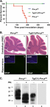
Scrapie-inoculated Tg(C1)/Prn-p0/0 mice do not develop clinical illness or histopathology, and do not accumulate protease-resistant PrP. A, survival was monitored in mice of the following genotypes after RML inoculation, with the number of animals indicated in parentheses: Prn-p0/0 (9), Tg(C1)/Prn-p0/0 (8), Prn-p+/+ (8). B, sections from the cerebellum (panels 1 and 2) or hippocampus (panels 3 and 4) from 365 day-old mice of the indicated genotypes were stained with hematoxylin and eosin or anti-GFAP antibody, respectively. Insets show hippocampal sections stained with DAPI to reveal cell nuclei. Scale bar in panel 4 (applicable to all panels) is 1 mm. C, Western blotting for protease-resistant PrP. Brain homogenates containing equivalent amounts of protein from mice of the indicated genotypes were treated with or without 20 μg/ml proteinase K for 1 h at 37 °C (+ and − lanes, respectively), and were subjected to Western blotting with anti-PrP antibody 6H4.
FIGURE 4.
FIGURE 4.
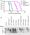
C1 prolongs survival time in mice expressing WT PrP. A, survival was monitored in mice of the following genotypes after scrapie inoculation, with the number of animals indicated in parentheses: Prn-p+/+ (8), Tg(C1)/Prn-p+/+ (8), Tga20+/0 (16), and Tg(C1)/Tga20+/0 (8). Significant differences were found between the following groups as determined by an unpaired student's t test: Tg(C1)/Prn-P+/+ versus Prn-P+/+ (p < 0.0001); Tg(C1)/Tga20+/0 versus Tga20+/0 (p < 0.0001). B, brain homogenates containing equal amounts of total protein from mice of the indicated genotypes were treated with or without PNGase F to remove N-linked oligosaccharides (+ and − lanes, respectively), and subjected to Western blotting with anti-PrP antibody D18. Filled and open arrowheads indicate the positions of cleavage products C1 and C2, respectively. Molecular size markers are given in kDa.
FIGURE 5.
FIGURE 5.
Effect of C1 on scrapie-induced pathology in terminally ill mice expressing WT PrP. Mice of the indicated genotypes were taken for histological analyses at the terminal stage of illness, with the age at sacrifice given in parentheses: Tg(C1)/Prn-p+/+ (233d), Prn-p+/+ (158d), Tg(C1)/Tga20+/0 (120d), Tga20+/0 (65d). Sections from the cerebellum were stained with hematoxylin and eosin (A–H) and sections from the hippocampus with anti-GFAP antibody (I–L). Areas within the cerebellar white matter outlined by the boxes in A–D are shown at higher magnification in E–H. Scale bars, 1 mm (A–D and I–L) and 50 μm (E–H).
FIGURE 6.
FIGURE 6.
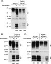
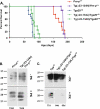
C1 inhibits accumulation of protease-resistant PrP in animals expressing WT PrP. Uninoculated or scrapie-inoculated mice (− and + lanes, respectively) of the indicated genotypes were sacrificed at the times shown below each lane. Brain homogenates containing equivalent amounts of protein were treated with (lower blots) or without (upper blots) 20 μg/ml proteinase K for 1 h at 37 °C, and were subjected to Western blotting with anti-PrP antibody 6H4. A shows samples from presymptomatic animals, and B and C show samples from terminally ill mice. Samples from two different mice at 180d, 80d, and 120d are shown. Actin is shown as a loading control in all panels.
FIGURE 7.
FIGURE 7.
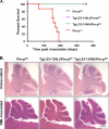
Scrapie-inoculated Tg(Δ23–134)/Prn-p0/0 mice do not develop clinical Illness or histopathology. A, survival was monitored in mice of the following genotypes after scrapie inoculation, with the number of animals indicated in parentheses: Prn-p0/0 (9), Tg(PrPΔ23–134L)/Prn-p0/0 (7), Tg(PrPΔ23–134H)/Prn-p0/0 (9), Prn-p+/+ (8). Data from RML-inoculated Prn-p0/0 and Prn-p+/+ mice shown in Fig. 4 are reproduced here to allow for direct comparison. B, mice were taken for histological analysis at 1 year of age, at which point they remained healthy. Cerebellar sections were stained with hematoxylin and eosin. Scale bar (applicable to all panels) is 1 mm.
FIGURE 8.
FIGURE 8.
Expression of PrP(Δ23–134) prolongs survival and reduces protease-resistant PrP. A, survival was monitored in mice of the following genotypes after scrapie inoculation, with the number of animals indicated in parentheses: Prn-p+/+ (8), Tg(Δ23–134H)/Prn-p+/+ (8), Tga20+/0 (16), Tg(Δ23–134L)/Tga20+/0 (9), and Tg(Δ23–134H)/Tga20+/0 (8). Significant differences were found between the following groups: Tg(Δ23–134H)/Prn-p+/+ versus Prn-p+/+ (p < 0.0001); and Tg(Δ23–134H)/Tga20+/0 versus Tga20+/0 (p < 0.0001) when analyzed either by Kruskal-Wallis one way analysis of variance with Dunn's Multiple Comparison Test, or by Students t test. C, animals of the indicated genotypes were sacrificed at the terminal stage of illness after scrapie inoculation. Brain homogenates containing equivalent amounts of protein were treated with (lower blots) or without (upper blots) 20 μg/ml proteinase K for 1 h at 37 °C, and were subjected to Western blotting with anti-PrP antibody 6H4.

Similar articles

Cited by

References

    1. Prusiner S. B. (1998) Proc. Natl. Acad. Sci. U.S.A. 95, 13363–13383 - PMC - PubMed
    1. Aguzzi A., Baumann F., Bremer J. (2008) Annu. Rev. Neurosci. 31, 439–477 - PubMed
    1. Pan K. M., Baldwin M., Nguyen J., Gasset M., Serban A., Groth D., Mehlhorn I., Huang Z., Fletterick R. J., Cohen F. E., Prusiner S. B. (1993) Proc. Natl. Acad. Sci. U.S.A. 90, 10962–10966 - PMC - PubMed
    1. Safar J., Roller P. P., Gajdusek D. C., Gibbs C. J., Jr. (1993) J. Biol. Chem. 268, 20276–20284 - PubMed
    1. Stahl N., Borchelt D. R., Hsiao K., Prusiner S. B. (1987) Cell 51, 229–240 - PubMed

Publication types