. 2011 Dec 15;589(Pt 24):6063-80.
doi: 10.1113/jphysiol.2011.215988. Epub 2011 Oct 24.

Dual role of junctin in the regulation of ryanodine receptors and calcium release in cardiac ventricular myocytes

Affiliations

Dual role of junctin in the regulation of ryanodine receptors and calcium release in cardiac ventricular myocytes

Beth A Altschafl et al. J Physiol. .

Abstract

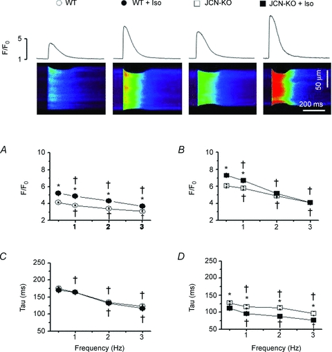
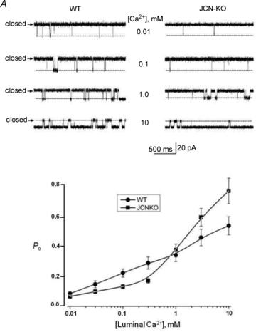
Junctin, a 26 kDa intra-sarcoplasmic reticulum (SR) protein, forms a quaternary complex with triadin, calsequestrin and the ryanodine receptor (RyR) at the junctional SR membrane. The physiological role for junctin in the luminal regulation of RyR Ca(2+) release remains unresolved, but it appears to be essential for proper cardiac function since ablation of junctin results in increased ventricular automaticity. Given that the junctin levels are severely reduced in human failing hearts, we performed an in-depth study of the mechanisms affecting intracellular Ca(2+) homeostasis in junctin-deficient cardiomyocytes. In concurrence with sparks, JCN-KO cardiomyocytes display increased Ca(2+) transient amplitude, resulting from increased SR [Ca(2+)] ([Ca(2+)](SR)). Junctin ablation appears to affect how RyRs 'sense' SR Ca(2+) load, resulting in decreased diastolic SR Ca(2+) leak despite an elevated [Ca(2+)](SR). Surprisingly, the β-adrenergic enhancement of [Ca(2+)](SR) reverses the decrease in RyR activity and leads to spontaneous Ca(2+) release, evidenced by the development of spontaneous aftercontractions. Single channel recordings of RyRs from WT and JCN-KO cardiac SR indicate that the absence of junctin produces a dual effect on the normally linear response of RyRs to luminal [Ca(2+)]: at low luminal [Ca(2+)] (<1 mmol l(-1)), junctin-devoid RyR channels are less responsive to luminal [Ca(2+)]; conversely, high luminal [Ca(2+)] turns them hypersensitive to this form of channel modulation. Thus, junctin produces complex effects on Ca(2+) sparks, transients, and leak, but the luminal [Ca(2+)]-dependent dual response of junctin-devoid RyRs demonstrates that junctin normally acts as an activator of RyR channels at low luminal [Ca(2+)], and as an inhibitor at high luminal [Ca(2+)]. Because the crossover occurs at a [Ca(2+)](SR) that is close to that present in resting cells, it is possible that the activator-inhibitor role of junctin may be exerted under periods of prevalent parasympathetic and sympathetic activity, respectively.


Figures

Figure 1
Figure 1. JCN-KO cardiomyocytes produce fewer but brighter Ca2+ sparks than WT cardiomyocytes
A and B, representative confocal line-scan images and three-dimensional Ca2+ sparks recorded in intact WT and JCN-KO cardiomyocytes, respectively. CG, bar graphs depicting Ca2+ spark frequency measured as the number of events per unit time and length, the maximal Ca2+ spark amplitude (F/F0), the average of full duration at half-maximal amplitude (FDHM), the average of full width at half-maximal amplitude (FWHM), and the time-to-peak (TTP) of Ca2+ spark amplitude in WT (blue bars) and JCN-KO (aqua bars) cells, respectively. ***P < 0.001. n = 1622 (WT) and 2743 (JCN-KO) Ca2+ sparks.
Figure 2
Figure 2. Isoproterenol perfusion attenuates the decreased Ca2+ spark frequency in JCN-KO cardiomyocytes
A and B, representative confocal line-scan images and three-dimensional Ca2+ sparks following the perfusion of 300 nmol l−1 Iso onto intact WT and JCN-KO cardiomyocytes, respectively. CG, bar graphs depicting Ca2+ spark frequency, amplitude, FDHM, FWHM and TTP, respectively, in Iso-perfused WT and JCN-KO cells. ***P < 0.001. n = 3837 (WT) and 2157 (JCN-KO) Ca2+ sparks.
Figure 3
Figure 3. JCN-KO cardiomyocytes display a significant increase in the amplitude of the intracellular Ca2+ transient and an isoproterenol-induced faster rate of decay
Representative confocal line scan images of Ca2+ transients elicited from cardiomyocytes field-stimulated at 0.5 Hz with the associated Ca2+ transient profiles (top panel). Line graphs depict the relationship between stimulation frequency and the amplitude of the Ca2+ transient (F/F0) (A and B) and the monoexponential decay of the Ca2+ transient (tau) (C and D). Open and filled symbols represent WT and JCN-KO cardiomyocytes, respectively. Circles and squares represent cardiomyocytes before and after 1 μmol l−1 Iso perfusion, respectively. †P < 0.05 vs. 0.5 Hz. *P < 0.05 JCN-KO vs. WT. n≥ 36 transients from ≥5 mouse hearts.
Figure 4
Figure 4. JCN-KO cardiomyocytes have a higher SR Ca2+ content than WT cardiomyocytes
A and B, representative line scan images and amplitude profiles of caffeine-induced Ca2+ transients elicited from WT and JCN-KO cardiomyocytes, respectively. C, bar graph depicting the amplitude (F/F0-caffeine) of the caffeine-elicited Ca2+ transients. *P < 0.05. n = 53 (WT) and 38 (JCN-KO) caffeine-induced Ca2+ transients.
Figure 5
Figure 5. The higher SR Ca2+ content in JCN-KO cardiomyocytes is potentially caused by a decrease in SR Ca2+ leak, which is reversed in the presence of isoproterenol
A and B, line graphs depicting the relationship between stimulation frequency and SR Ca2+ leak in the absence and presence of 1 μmol l−1 Iso, respectively. C and D, bar graphs depicting the combined SR Ca2+ leak in the absence and presence of 1 μmol l−1 Iso, respectively. *P < 0.05. n≥ 6 measurements per pacing frequency from 4 hearts. E, bar graphs depicting the percentage of WT (n = 5 of 87) and JCN-KO (n = 32 of 66) cardiomyocytes that developed spontaneous aftercontractions. Data were analysed using a Chi2 test. *P < 0.05. F and G, WT and JCN-KO cardiomyocytes were paced for 2–3 s at 3 Hz in the presence of 1 μmol l−1 Iso, then pacing was stopped to image spontaneous Ca2+ release events. Representative Ca2+ transient profiles (top traces), line scan images (middle images), and cell shortening profiles (bottom traces) from a WT and JCN-KO cardiomyocyte.
Figure 6
Figure 6. Effect of junctin removal on single channel RyR2 activity is dependent on the luminal Ca2+ concentration
A, single RyR channels recorded from WT and JCN-KO cardiac microsomes reconstituted into a planar lipid bilayer with 0.01–10 mmol l−1 Ca2+ bathing the trans (luminal) side of the channel. B, data points depict the relationship between luminal [Ca2+] and the open probability (Po) of the RyR2 channel as a function of luminal [Ca2+]. Circles and squares represent WT and JCN-KO single channel data, respectively. Data points are joined by a line with no theoretical function and correspond to the mean ± SEM for n = 8 and 6 channels (WT and JCN-KO, respectively).
Figure 7
Figure 7. Junctin and RyR2 interacting domains
A, diagram depicting the junctin and RyR2 interacting domains probed in this study. B, MBP–junctin fusion peptides separated by electrophoresis were transferred to membranes and blot-overlaid with GST–RyR2-A and GST–RyR2-D. C, GST–RyR2 fusion peptides separated by electrophoresis were transferred to membrane and blot-overlaid with MBP–junctin iso(1), MBP–junctin-A and MBP–junctin-D.
Figure 8
Figure 8. Proposed model for junctin effect on RyR2 channel activity
Two binding sites for junctin (labelled ‘1’ and ‘2’ in the WT cartoons) on the luminal side of the cardiac RyR2 may mediate the dual role of junctin described in this study. Arrows on top of RyR2 represent Po of RyR2, Ca2+ spark frequency, or SR Ca2+ leak during diastole. Notice the variable thickness of the arrows. Left and right cartoon in each panel represent the SR Ca2+ loading during basal conditions and during β-adrenergic stimulation, respectively. See text for more details.

References

    1. Altschafl BA, Beutner G, Sharma VK, Sheu SS, Valdivia HH. The mitochondrial ryanodine receptor in rat heart: A pharmaco-kinetic profile. Biochim Biophys Acta. 2007;1768:1784–1795. - PubMed
    1. Benkusky NA, Weber CS, Scherman JA, Farrell EF, Hacker TA, Powers PA, Valdivia HH. Intact β-adrenergic response and unaltered progression towards heart failure in mice with genetic ablation of a major PKA phosphorylation site in the cardiac ryanodine receptor. Circ Res. 2007;101:819–829. - PubMed
    1. Bers DM. Cardiac excitation-contraction coupling. Nature. 2002;415:198–205. - PubMed
    1. Capes EM, Loaiza R, Valdivia HH. Ryanodine Receptors. Skelet Muscle. 2011;1:18–31. - PMC - PubMed
    1. Cheng H, Lederer WJ. Calcium sparks. Physiol Rev. 2008;88:1491–1545. - PubMed

Publication types

MeSH terms

LinkOut - more resources