. 2011 Nov 8;108(45):18530-5.
doi: 10.1073/pnas.1106706108. Epub 2011 Oct 24.

CLUMPED CHLOROPLASTS 1 is required for plastid separation in Arabidopsis

Affiliations

CLUMPED CHLOROPLASTS 1 is required for plastid separation in Arabidopsis

Yue Yang et al. Proc Natl Acad Sci U S A. .

Abstract

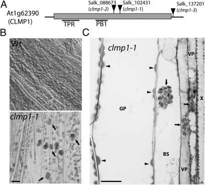
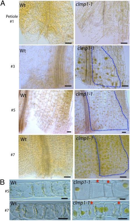
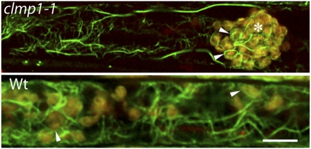
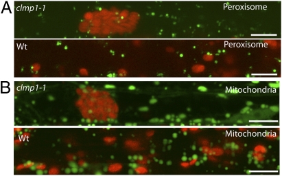
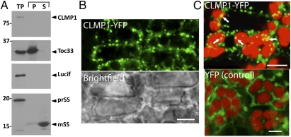
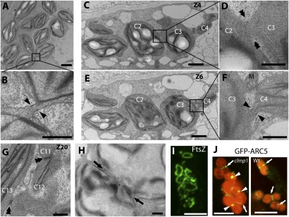
We identified an Arabidopsis thaliana mutant, clumped chloroplasts 1 (clmp1), in which disruption of a gene of unknown function causes chloroplasts to cluster instead of being distributed throughout the cytoplasm. The phenotype affects chloroplasts and nongreen plastids in multiple organs and cell types, but is detectable only at certain developmental stages. In young leaf petioles of clmp1, where clustering is prevalent, cells lacking chloroplasts are detected, suggesting impaired chloroplast partitioning during mitosis. Although organelle distribution and partitioning are actin-dependent in plants, the actin cytoskeleton in clmp1 is indistinguishable from that in WT, and peroxisomes and mitochondria are distributed normally. A CLMP1-YFP fusion protein that complements clmp1 localizes to discrete foci in the cytoplasm, most of which colocalize with the cell periphery or with chloroplasts. Ultrastructural analysis revealed that chloroplasts within clmp1 clusters are held together by membranous connections, including thin isthmi characteristic of late-stage chloroplast division. This finding suggests that constriction of dividing chloroplasts proceeds normally in clmp1, but separation is impaired. Consistently, chloroplast size and number, as well as positioning of the plastid division proteins FtsZ and ARC5/DRP5B, are unaffected in clmp1, indicating that loss of CLMP1-mediated chloroplast separation does not prevent otherwise normal division. CLMP1-like sequences are unique to green algae and land plants, and the CLMP1 sequence suggests that it functions through protein-protein interactions. Our studies identify a unique class of proteins required for plastid separation after the constriction stage of plastid division and indicate that CLMP1 activity is also required for plastid distribution and partitioning during cell division.


Conflict of interest statement

The authors declare no conflict of interest.

Figures

Fig. 1.
Fig. 1.
Identification of CLMP1. (A) Gene structure and T-DNA insertion mutations. The box indicates the exon; the thin black lines represent UTRs. Positions of the predicted TPR and PB1 domains are indicated. (B) Chloroplast phenotype in WT and clmp1-1. Arrows indicate clumped chloroplasts. (C) Light micrograph of a petiole section from clmp1-1. The WT control is shown in Fig. S1A. Arrows indicate clumped chloroplasts; arrowheads, dispersed chloroplasts. X, xylem; VP, vascular parenchyma cell; BS, bundle sheath cell; GP, ground parenchyma cell. (Scale bars: 20 μm.)
Fig. 2.
Fig. 2.
More detailed characterization of clmp1. (A) Whole-mount differential interference contrast images of the petiole base from leaves 1, 3, 5, and 7 (old to young) of WT and clmp1-1. Dotted lines outline regions in which clumped chloroplasts are observed within half a petiole. (Scale bars: 50 μm.) (B) Ground parenchyma cells isolated from petioles of leaves 5 and 7 of WT and clmp1-1. Cells were from regions corresponding to outlined areas of leaves 5 and 7 in A. Red stars indicate cells lacking chloroplasts. (Scale bars: 20 μm.)
Fig. 3.
Fig. 3.
Alexa Fluor 488-phalloidin staining (green) of actin in VP cells in clmp1-1 and WT. Chlorophyll autofluorescence is indicated in red. (Scale bar: 10 μm.) Asterisk denotes clumped chloroplasts; arrowheads indicate chloroplast-associated actin filaments.
Fig. 4.
Fig. 4.
Distribution of peroxisomes (A) and mitochondria (B) in the VP cells of clmp1-1 and WT. Peroxisomes and mitochondria are in green; chlorophyll autofluorescence is in red. Images were superimposed from three Z-section images. (Scale bars: 10 μm.)
Fig. 5.
Fig. 5.
CLMP1 localization. (A) Chloroplast import assay of CLMP1 and controls. TP, translation product; P, chloroplast membrane fraction; S, chloroplast soluble fraction; PrSS, SS precursor; mSS, mature SS. Positions of molecular mass markers (in kDa) are shown at the left. (B) CLMP1-YFP localizes to foci near the cell periphery in complemented clmp1-1 plants. A brightfield image of the same sample is shown below the fluorescence image. (C) Some CLMP1-YFP foci colocalize with chloroplasts (arrows). YFP localization in WT is shown as a control. YFP fluorescence is indicated in green; chlorophyll autofluorescence is in red. (Scale bars: 5 μm.)
Fig. 6.
Fig. 6.
Structure of clumped chloroplasts in clmp1. (A–H) Transmission electron micrographs of chloroplasts within clumps. CG are selected images from serial sections of a single chloroplast cluster shown more completely in Figs. S7 and S9. Chloroplasts with the same number in CG and Fig. S7 (arbitrarily numbered C1–C13) represent the same chloroplast viewed in different serial sections. A, B, and H are from a different cluster. B, D, and F are magnified views of the boxes in A, C, and E, respectively. Black arrowheads indicate presumed PD rings at the edges of isthmi. Double arrowheads indicate chloroplast connections that are disorganized in appearance. Black arrows indicate chloroplast constriction sites. M, mitochondrion. (Scale bars: 2 μm in A, C, and E; 500 nm in B, D, F, G, and H.) (I) FtsZ immunolocalization in clmp1. (Scale bar: 5 μm.) (J) GFP-ARC5 localization in clmp1 and WT. (Scale bar: 5 μm.)

References

    1. Wada M, Suetsugu N. Plant organelle positioning. Curr Opin Plant Biol. 2004;7:626–631. - PubMed
    1. Miyagishima SY, Kabeya Y. Chloroplast division: Squeezing the photosynthetic captive. Curr Opin Microbiol. 2010;13:738–746. - PubMed
    1. Osteryoung KW, Nunnari J. The division of endosymbiotic organelles. Science. 2003;302:1698–1704. - PubMed
    1. Kuroiwa T, et al. Vesicle, mitochondrial, and plastid division machineries with emphasis on dynamin and electron-dense rings. Int Rev Cell Mol Biol. 2008;271:97–152. - PubMed
    1. Vitha S, McAndrew RS, Osteryoung KW. FtsZ ring formation at the chloroplast division site in plants. J Cell Biol. 2001;153:111–120. - PMC - PubMed

Publication types

MeSH terms

Substances