. 2011 Sep;3(9):873-88.
doi: 10.18632/aging.100391.

Replicative senescence of mesenchymal stem cells causes DNA-methylation changes which correlate with repressive histone marks

Affiliations

Replicative senescence of mesenchymal stem cells causes DNA-methylation changes which correlate with repressive histone marks

Anne Schellenberg et al. Aging (Albany NY). 2011 Sep.

Abstract

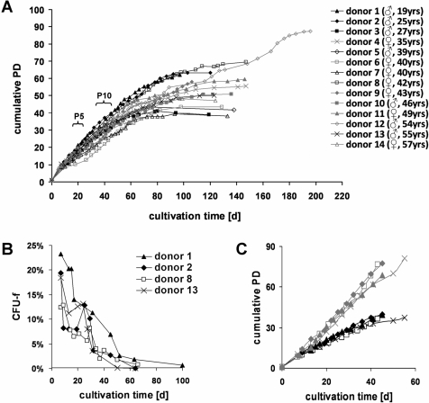
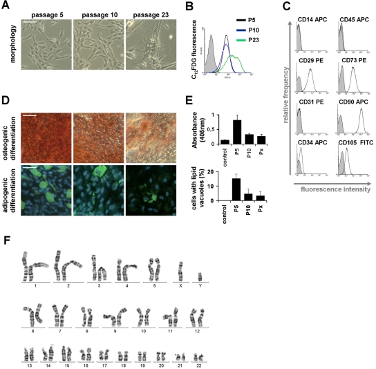
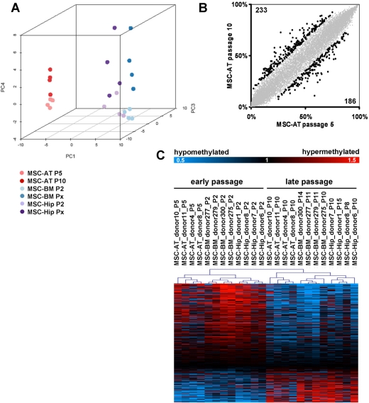
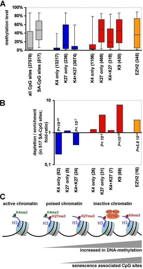
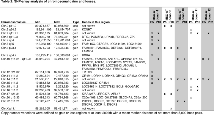
Cells in culture undergo replicative senescence. In this study, we analyzed functional, genetic and epigenetic sequels of long-term culture in human mesenchymal stem cells (MSC). Already within early passages the fibroblastoid colony-forming unit (CFU-f) frequency and the differentiation potential of MSC declined significantly. Relevant chromosomal aberrations were not detected by karyotyping and SNP-microarrays. Subsequently, we have compared DNA-methylation profiles with the Infinium HumanMethylation27 Bead Array and the profiles differed markedly in MSC derived from adipose tissue and bone marrow. Notably, all MSC revealed highly consistent senescence-associated modifications at specific CpG sites. These DNA-methylation changes correlated with histone marks of previously published data sets, such as trimethylation of H3K9, H3K27 and EZH2 targets. Taken together, culture expansion of MSC has profound functional implications - these are hardly reflected by genomic instability but they are associated with highly reproducible DNA-methylation changes which correlate with repressive histone marks. Therefore replicative senescence seems to be epigenetically controlled.


Conflict of interest statement

The authors of this manuscript have no conflict of interest to declare.

Figures

Figure 1
Figure 1. Long-term growth curves of MSC
Mesenchymal stem cells were isolated from human adipose tissue of 14 donors and culture expanded until the cells reached a senescent state. Every cell passage is indicated by a point and the number of cumulative population doublings (PD) was calculated based on the ratio of cells seeded versus cells harvested per passage (A). In parallel, MSC were seeded in limiting dilution to determine the fibroblastoid colony forming unit (CFU-f) frequency for subsequent passages (B). To account for the fact, that the progeny of each passage is based on a decreasing percentage of highly proliferative cells, we have recalculated long-term growth curves on the basis of the number of CFU-f seeded versus cells harvested per passages (C; black: conventional PD; grey: CFU-f-adopted growth curves).
Figure 2
Figure 2. Changes of MSC during culture expansion
Senescent MSC acquire a typical large and flat morphology, whereas no differences were observed at passage 5 and passage 10 (A). Expression of the senescence-associated β-galactosidase was detected with the fluorogenic substrate C12FDG and this biomarker for replicative senescence was positive in senescent passages but not in passage 5 and passage 10 (B). All MSC preparations displayed the typical immunophenotype (C) and could be induced towards osteogenic and adipogenic lineage (D). However, this in vitro differentiation potential decayed already between passage 5 and passage 10 (E; control is without induction medium; Px represents the corresponding senescent passage; all data are presented as mean ± SD; n = 4). A normal karyogram of MSC (after treatment with trypsin and Giemsa)is exemplarily presented (F). (scale bars = 100μm).
Table 2
Table 2
Figure 3
Figure 3. Senescence-associated modifications in the DNA-methylation pattern
DNA-methylation profiles were analyzed with the HumanMethylation27 BeadChip microarray which represents 27,578 unique CpG sites. MSC derived from adipose tissue (MSC-AT) were compared with those derived from bone marrow, which was either aspirated from the iliac crest (MSC-BM) or taken from the caput femuris upon hip replacement (MSC-Hip). Unsupervised principal component analysis (PCA) clearly separated DNA-methylation profiles according to the tissue of origin in the first dimension (PC1), whereas the forth component (PC4) discerned early and late passage (A). Scatterplot comparison of passage 5 and passage 10 in MSC-AT revealed that 233 CpG sites are more than 15% hyper-methylated whereas 186 CpG sites are more than 15% hypo-methylated at passage 10 (B). Significance Analysis of Microarray (SAM) was used to select 517 senescence-associated CpG sites (FDR = 4.8%) and these are presented as a heatmap (C; data were divided by the mean of each row for graphical presentation).
Figure 4
Figure 4. Senescence-associated DNA-methylation changes correlate with repressive histone marks
DNA-methylation profiles of MSC were compared with datasets for H3K4me3 and H3K27me3 in human ESC [34] (indicated in blue); H3K4me3, H3K9me3 and H3K27me3 in human MSC-AT [35] (red) and targets of the Enhancer of Zeste Homolog 2 (EZH2) in human MSC-BM [36] (orange). The overall DNA-methylation level of all 27,578 CpG sites on the microarray was much lower in comparison to the 517 senescence-associated (SA) CpG sites and to those which have been assigned for H3K27me3, H3K9me3 and targets of EZH2 (A; Box plots represent the 25th and 75th percentile for each subset and whiskers show the 5% and 95% percentiles). Notably, the 517 senescence-associated DNA-methylation changes were significantly enriched in regions with H3K27me3, H3K9me3 and EZH2 targets (B). The graphic illustrates that repressive histone marks are associated with higher levels of DNA-methylation and that senescence-associated modifications are enriched in these regions (C).

References

    1. Hayflick L., Moorhead P.S. The serial cultivation of human diploid cell strains. Exp Cell Res. 1961;25:585–621. - PubMed
    1. Greider C.W., Blackburn E.H. Identification of a specific telomere terminal transferase activity in Tetrahymena extracts. Cell. 1985;43:405–413. - PubMed
    1. Di Donna S., Mamchaoui K., Cooper R.N., Seigneurin-Venin S., Tremblay J., Butler-Browne G.S., Mouly V. Telomerase can extend the proliferative capacity of human myoblasts, but does not lead to their immortalization. Mol Cancer Res. 2003;1:643–653. - PubMed
    1. Masutomi K., Yu E.Y., Khurts S., Ben-Porath I., Currier J.L., Metz G.B., Brooks M.W., Kaneko S., Murakami S., DeCaprio J.A., Weinberg R.A., Stewart S.A., Hahn W.C. Telomerase maintains telomere structure in normal human cells. Cell. 2003;114:241–253. - PubMed
    1. Chen Q., Fischer A., Reagan J.D., Yan L.J., Ames B.N. Oxidative DNA damage and senescence of human diploid fibroblast cells. Proc Natl Acad Sci U S A. 1995;92:4337–4341. - PMC - PubMed

Publication types

MeSH terms