Retreatodontics in maxillary lateral incisor with supernumerary root
- PMID: 22025843
- PMCID: PMC3198569
- DOI: 10.4103/0972-0707.85827
Retreatodontics in maxillary lateral incisor with supernumerary root
Abstract
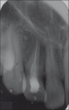
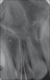
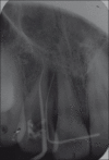
Familiarity with the intricacies and variations of root canal morphology is essential for successful endodontic treatment. Maxillary central and lateral incisors are known to be single rooted with one canal. This case report describes endodontic retreatment of maxillary lateral incisors with two root canals, one of which was missed during the initial treatment.
Keywords: Endodontic retreatment; extra canals; maxillary lateral incisor; supernumerary root.
Conflict of interest statement
Figures


References
-
- Pecora JD, Santana SV. Maxillary lateral incisor with two roots-case report. Braz Dent J. 1992;2:151–3. - PubMed
-
- Walvekar SV, Behbehani JM. Three root canals and dens formation in a maxillary lateral incisor: A case report. J Endod. 1997;23:185–6. - PubMed
-
- Neville BW, Damm DD, Allen CM, Bouquot JE. Oral and maxillofacial pathology. 2nd ed. Philadelphia: W.B. Saunders; 2002. p. 88.
-
- Kelly JR. Birooted primary canines. Oral Surg Oral Med Oral Pathol. 1978;46:872. - PubMed
-
- Vertucci FJ. Root canal anatomy of the human permanent teeth. Oral Surg Oral Med Oral Pathol. 1984;58:589–99. - PubMed

