

Congenital megalocornea with zonular weakness and childhood lens-related secondary glaucoma - a distinct phenotype caused by recessive LTBP2 mutations
- PMID: 22025892
- PMCID: PMC3198484
Congenital megalocornea with zonular weakness and childhood lens-related secondary glaucoma - a distinct phenotype caused by recessive LTBP2 mutations
Abstract
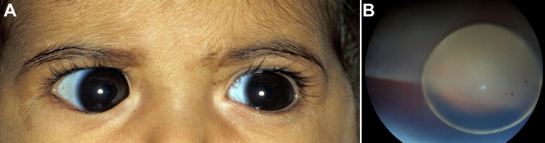
Purpose: To clinically and genetically characterize a distinct phenotype of congenital megalocornea (horizontal corneal diameter ≥13 mm) with secondary glaucoma from spherophakia and/or ectopia lentis during childhood in affected Saudi families.
Methods: Clinical exam, homozygosity scan, and candidate gene analysis.
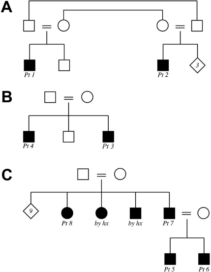
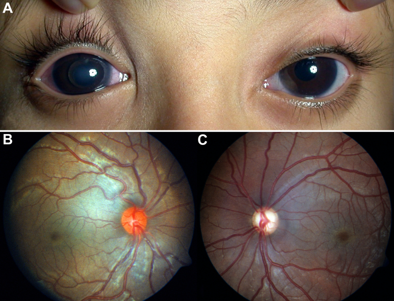
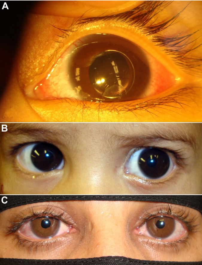
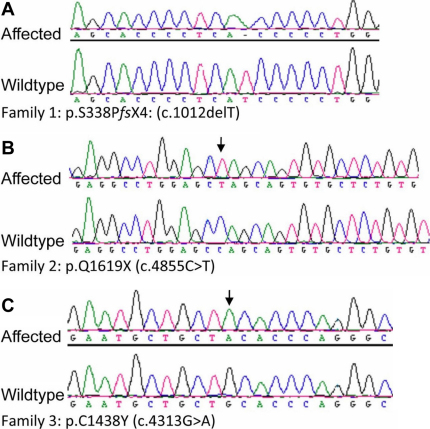
Results: From 2005 to 2010, eight affected individuals from three consanguineous families were identified. In addition to congenital megalocornea, affected children presented with secondary glaucoma from spherophakia and/or ectopia lentis. One member from each family developed spontaneous complete crystalline lens dislocation into the anterior chamber with associated acute glaucoma during early childhood. Older individuals had phenotypes that would have suggested prior uncontrolled primary congenital/infantile glaucoma had past ophthalmic and/or family histories not been available. Homozygosity mapping performed for the first two families suggested the candidate gene latent transforming growth factor-beta-binding protein 2 (LTBP2), which when sequenced revealed a novel homozgyous mutation that segregated with the phenotype in each family (p.S338PfsX4 [c.1012delT], p.Q1619X[(c.4855C>T]). LTBP2 sequencing in the third family revealed a third novel homozygous mutation (p.C1438Y [c.4313G>A]).
Conclusions: Congenital megalocornea with childhood secondary glaucoma from spherophakia and/or ectopia lentis is a distinct condition caused by recessive LTBP2 mutations that needs to be distinguished from buphthalmos secondary to primary congenital/infantile glaucoma because typical initial surgical treatment is lens removal in the former and angle surgery in the latter. Complete dislocation of the crystalline lens into the anterior chamber during early childhood can occur in young children with this unique phenotype.
Figures
References
-
- Ho CL, Walton DS. Primary congenital glaucoma: 2004 update. J Pediatr Ophthalmol Strabismus. 2004;41:271–88. - PubMed
-
- Khan AO. Genetics of primary glaucoma. Curr Opin Ophthalmol. 2011;22:347–55. - PubMed
-
- Al-Harthi E, Al-Shahwan S, Al-Turkmani S, Khan AO. Retinal detachment and congenital glaucoma. Ophthalmology. 2007;114:1590–1. - PubMed
-
- Khan AO. A surgical approach to paediatric glaucoma. Ophthalmology International. 2009;4:77–81.
-
- Khan AO. Conditions that can be mistaken for childhood glaucoma. Ophthalmic Genet. 2011;32:129–37. - PubMed
Publication types
MeSH terms
Substances
LinkOut - more resources
Full Text Sources
Medical
Molecular Biology Databases
Miscellaneous