. 2008 Sep 24:2:43-74.
doi: 10.4137/mri.s1131.

DANCING WITH THE ELECTRONS: TIME-DOMAIN AND CW IN VIVO EPR IMAGING

Affiliations

DANCING WITH THE ELECTRONS: TIME-DOMAIN AND CW IN VIVO EPR IMAGING

Sankaran Subramanian et al. Magn Reson Insights. .

Abstract

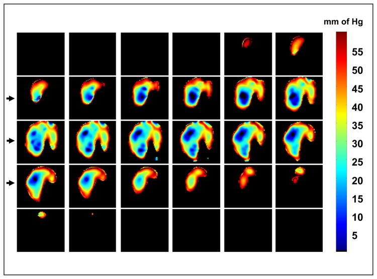
The progress in the development of imaging the distribution of unpaired electrons in living systems and the functional and the potential diagnostic dimensions of such an imaging process, using Electron Paramagnetic Resonance Imaging (EPRI), is traced from its origins with emphasis on our own work. The importance of EPR imaging stems from the fact that many paramagnetic probes show oxygen dependent spectral broadening. Assessment of in vivo oxygen concentration is an important factor in radiation oncology in treatment-planning and monitoring treatment-outcome. The emergence of narrow-line trairylmethyl based, bio-compatible spin probes has enabled the development of radiofrequency time-domain EPRI. Spectral information in time-domain EPRI can be achieved by generating a time sequence of T(2)* or T(2) weighted images. Progress in CW imaging has led to the use of rotating gradients, more recently rapid scan with direct detection, and a combination of all the three. Very low field MRI employing Dynamic Nuclear polarization (Overhauser effect) is also employed for monitoring tumor hypoxia, and re-oxygenation in vivo. We have also been working on the co-registration of MRI and time domain EPRI on mouse tumor models at 300 MHz using a specially designed resonator assembly. The mapping of the unpaired electron distribution and unraveling the spectral characteristics by using magnetic resonance in presence of stationary and rotating gradients in indeed 'dancing with the (unpaired) electrons', metaphorically speaking.


Figures

Fig. 1
Fig. 1
The molecular structure of the symmetric triarylmethyl derivatives, Oxo31 and Oxo63 used for CW and time-domain EPR imaging in our laboratory.
Fig. 2
Fig. 2
Concept of spectral-spatial imaging. Obtaining several sets of projections form an object with gradually increasing gradients, and rescaling the projections to same size generates views of the object in a pseudo spatial-spectral dimension, leading to the separation of the spectral information from the spatial information.
Fig. 3
Fig. 3
Simplified schematic of stepped-field rotating gradient 2D CW EPR. The field is stepped through 8 points (top) and for each field point the gradients rotated in a plane by one full cycle. In this simplified example, the n points collected per gradient cycle are sub-sampled to 8 points to produce the Bθ matrix (Fig. 4) column-wise. By carefully adjusting the field scan rate, the gradient rotating frequency and the sampling speed one can optimize the image resolution.
Fig. 4
Fig. 4
Elements of the Bθ matrix that represents the projections obtained at a constant gradient as a function of the orientation of the gradient vector. Elements are collected row-wise in conventional EPR imaging and column-wise in rotating-gradient stepped-field data collection. The large horizontal arrow represents the conventional way of collection projections at constant gradient vector at a given orientation, and then changing the orientation sequentially. The big vertical arrow represents the stepped-gradient-rotating-gradient approach. The big slanted arrow represents the present method that corresponds to the simultaneous application of the field sweep field and rotating gradients.
Fig. 5
Fig. 5
The Bθ projection matrix. The conventional projection data collection in presence of constant orientation of gradients is represented by the rows of the matrix. The stepped-field, rotating gradient method is represented by the columns. The simultaneous rapid-scan in presence of rotating gradients modality is represented by directions parallel to the diagonal (or anti-diagonal) of the matrix.
Fig. 6
Fig. 6
Simplified schematics of the Rapid scan rotating gradient CW EPR Imager.
Fig. 7
Fig. 7
Direct detected absorption EPR signals of a phantom sample in the absence of gradients. The top sinusoid represents the rapid scan sweep (1.2 kHz), and the two high frequency sinusoids (blue and red, 4.8 kHz) represent the x and z gradients, which together provide the rotating gradient in the xz plane. The left side of the dotted rectangle represents the start of the trigger. 64 samples at 600 kS/s were collected for each gradient phase setting, giving rise to one downfield scan and one up field scan. Acquisition time per projection is 208 μs.
Fig. 8
Fig. 8
Conventional sinogram from a 2-tube phantom (on the left) and the corresponding filtered back-projected image (left column I) Skewed pseudo sonogram and the corresponding pseudo image obtained from the raw data when projections were collected with simultaneous sweep and rotating gradients (middle column II). Reshuffling the matrix leads to the correct sinogram and the expected image (last column III).
Fig. 9
Fig. 9
Schematics of Single Point Imaging (SPI). The FIDs are collected in presence of phase encoding gradients and the phase modulation on a given single time point after a delay τ leads to a response that is analogous to the gradient recalled echo in MRI. The gradients are ramped from positive maximum to negative maximum value in incremental steps, and the resulting phase modulation leads to amplitude modulation, encoding the spatial location of spins. Fourier transformation of the echo leads to the image. The gradients can be looped to generate 1, 2 or 3D k-space.
Fig. 10
Fig. 10
Schematics of the multigradient SPI experiment to evaluate T2* decay**. A series of interleaved SPI experiments at different gradients allows selecting single point images at different delays with almost identical resolution. At higher gradients the decay rate is faster and SNR is better at short delays, and at lower gradients the decay rate is slower and SNR is comparable at larger delays. Combining the results from multigradient experiements the decay rate when the FOV’s are almost the same leads to reproducible single exponential T2* and apparent line widths.
Fig. 11
Fig. 11
Schematics of the OMRI pulse sequence which is quite similar to the MRI gradient recalled echo sequence except for the electron pre-saturation pulse indicated by EPR. Two gradient echo images are sequentially measured for two different power levels in order to evaluate the two unknowns, the probe concentration and the in vivo pO2. What is not shown is that EPR irradiation is carried out at 226 MHz at 8.1 mT and just before MRI at 640 kHz, the field is ramped up to 15 mT.
Fig. 12
Fig. 12
3D SPI image of a Spiral phantom. Schematics of the glass spiral tubing filled with 2 mM Oxo63 in saline is shown on the left. The inside diameter of the glass tube was 1.4 mm and the capacity of the spiral was around 450 μL. The gradient increment was 0.3 Gauss/cm and the maximum gradient was 1.5 Gauss/cm. The data matrix size was 11 × 11 × 11 (1331 single points) and the size of the image matrix was 64 × 64 × 64. The 3D rendering at a few orientations shown on the left was carried out using the software Voxelview® in a silicon graphics Indigo2 workstation. The quality of the image is very good and dimensions matched well with those of the phantom.
Fig. 13
Fig. 13
Left: The linearity of the T2* based line width versus the dissolved oxygen concentartion for a 4-tube phantom with 2 mM Oxo63 saturated respectively with 0, 1, 2.5 and 5% oxygen, obtained from a 3-gradient, 21 × 21 × 21 steps oxymetric SPI 3D imaging data. Right: The corresponding color-coded oxygen images clearly distinguishing small differences in oxygen concentration.
Fig. 14
Fig. 14
Left: The cartoon of a C3H mouse with its tumor-bearing and normal legs placed inside a 25 × 25 mm cylindrical resonator, separated by a Lucite partition. Right: After the mouse was infused with 75 μL 100 mM Oxo63 (approx. ~3 mM probe concentration in the blood), 3D SPI images were obtained using a maximum gradient of 1.2 G/cm, 21 × 21 × 21 gradient steps and 2000 averages in about 2.5 min. Surface rendered 3D image showing clearly the excellent image of the uniform outer distribution of the spin, the location of the tumor and the bladder.
Fig. 15
Fig. 15
Axial cut-away views of spin distribution in normal and SCC tumor-bearing legs of a C3H mouse from 3D single point EPR image data shown above. 2D slices shown at 2.5 mm intervals. It can be clearly seen the tumor is preferentially perfused with blood compared to normal leg.
Fig. 16
Fig. 16
Coronal slices (1.1 mm thick) of oxygen distribution through the normal and SCC tumor-bearing legs of a C3H mouse derived from a 3-gradient 3D SPI image data based on T2*. The total measuring time was 7.5 min. The hypoxic zones with near-zero pO2 on the tumor leg, and the relatively uniformly oxygenated normal leg can be clearly monitored. The heterogeneity of oxygen distribution in the tumor leg is also clearly seen.
Fig. 17
Fig. 17
A. Coronal image (5 mm slice) of a C3H mouse with SCC tumor implanted in one of the legs taken at 640 kHz (low field MRI) with Overhauser enhancement. The kidneys and the tumor areas are well perfused by the spin probe and show good enhancement. B. Calculated spin probe image showing up to 3mM concentration of trityl in the tumor. C. Calculated oxygen image showing close to 80 mm of pO2 in the kidneys and very low (<10 mm) pO2 in the tumor zone. D. Zoomed-in image of the tumor-bearing leg showing large areas of near-zero (hypoxic) pO2. E. Oxygen map of the same slice as D, but the when the mouse is allowed to breathe Crabogen (95% O2/5% CO2) clearly showing increased oxygenation.
Fig. 18
Fig. 18
Schemaitic representation of EPRI-MRI coregistration experiment. A. The 300 MHz EPRI hardware. B. The resonator holder assembly that fits into the EPR magnet during EPR imaging. C. The MRI gantry that co-axially holds the EPR-resonator assembly and fits in the 7T MRI magnet for performing various functional MR imaging. Immediately following EPRI the mouse along with EPR resonator is fixed on to the MRI gantry without the need to move the mouse. A switch in the resonator circuit allows going from low Q (15 for EPRI) to high Q (200 for MRI) D. The 7T MR spectrometer (Bruker Biospin).
Fig. 19
Fig. 19
EPR oxygen imaging of normal muscle in live mice. EPRI method allows the pO2 map from deep in tissue of healthy mouse to be obtained. The anatomic image from MRI (A) of the lower body of a healthy mouse (without tumor bearing) and corresponding pO2 map from EPRI (B) showed that the normal muscle region had relatively homogeneous pO2 distribution. The anatomic image (C) and pO2 image (D) were also obtained from the contra-lateral normal leg of a SCC tumor-bearing mouse, and compared with that of a healthy mouse. (E) There was no significant difference in pO2 between normal muscle tissues with or without tumor bearing as opposed to the significant, lower pO2 in tumor region of the SCC mouse.
Fig. 20
Fig. 20
Co-registered MRI and EPR images. The left column shows coronal slices (5 mm) through the SCC tumor-bearing and normal mouse legs from T2-weighted MRI. The middle column shows the blood volume (%) image (MRI) derived from difference in GE image intensities before and after the injection of USPIO (ultra small super paramagnetic iron oxide). The right column shows the pO2 map obtained from multi-gradient EPR SPI. The trend in the oxygenation (modulated by allowing the mouse to breathe gases with different percentage of oxygen, 95%, 10% and 20%) is faithfully revealed by the EPR oxygen maps and shows a parallel to the blood-volume images from MRI.

Similar articles

  • 3-Carboxy-2,2,5,5-tetramethyl-pyrrolidinyl-N-oxyl.
    Zhang H. Zhang H. 2008 Apr 30 [updated 2008 Jun 9]. In: Molecular Imaging and Contrast Agent Database (MICAD) [Internet]. Bethesda (MD): National Center for Biotechnology Information (US); 2004–2013. 2008 Apr 30 [updated 2008 Jun 9]. In: Molecular Imaging and Contrast Agent Database (MICAD) [Internet]. Bethesda (MD): National Center for Biotechnology Information (US); 2004–2013. PMID: 20641530 Free Books & Documents. Review.
  • 15N-Labeled 4-oxo-2,2,6,6-tetramethyl-piperidine-1-oxyl.
    Zhang H. Zhang H. 2008 Apr 30 [updated 2008 Jun 9]. In: Molecular Imaging and Contrast Agent Database (MICAD) [Internet]. Bethesda (MD): National Center for Biotechnology Information (US); 2004–2013. 2008 Apr 30 [updated 2008 Jun 9]. In: Molecular Imaging and Contrast Agent Database (MICAD) [Internet]. Bethesda (MD): National Center for Biotechnology Information (US); 2004–2013. PMID: 20641553 Free Books & Documents. Review.
  • 3-Carbamoyl-2,2,5,5-tetramethyl-1-pyrrolidinyl-N-oxyl.
    Zhang H. Zhang H. 2008 Apr 30 [updated 2008 Jun 9]. In: Molecular Imaging and Contrast Agent Database (MICAD) [Internet]. Bethesda (MD): National Center for Biotechnology Information (US); 2004–2013. 2008 Apr 30 [updated 2008 Jun 9]. In: Molecular Imaging and Contrast Agent Database (MICAD) [Internet]. Bethesda (MD): National Center for Biotechnology Information (US); 2004–2013. PMID: 20641525 Free Books & Documents. Review.
  • In vivo imaging of a stable paramagnetic probe by pulsed-radiofrequency electron paramagnetic resonance spectroscopy.
    Murugesan R, Cook JA, Devasahayam N, Afeworki M, Subramanian S, Tschudin R, Larsen JA, Mitchell JB, Russo A, Krishna MC. Murugesan R, et al. Magn Reson Med. 1997 Sep;38(3):409-14. doi: 10.1002/mrm.1910380309. Magn Reson Med. 1997. PMID: 9339442
  • EPR and Related Magnetic Resonance Imaging Techniques in Cancer Research.
    Takakusagi Y, Kobayashi R, Saito K, Kishimoto S, Krishna MC, Murugesan R, Matsumoto KI. Takakusagi Y, et al. Metabolites. 2023 Jan 1;13(1):69. doi: 10.3390/metabo13010069. Metabolites. 2023. PMID: 36676994 Free PMC article. Review.

Cited by

References

    1. Alecci M, Dellapenna S, Sotgui A, et al. Electron-Paramagnetic Resonance Spectrometer for 3-Dimensional Invivo Imaging at Very Low-Frequency. Rev Sci Instrum. 1992;63:4263–4270.
    1. Alecci M, Brivati JA, Placidi G, et al. A submicrosecond resonator and receiver system for pulsed magnetic resonance with large samples. J Magn Reson. 1998;132:162–166.
    1. Ardenkjaer-Larsen JH, Laursen I, Leunbach I, et al. EPR and DNP properties of certain novel single electron contrast agents intended for oximetric imaging. J Magn Reson. 1998;133:1–12. - PubMed
    1. Balcom B, Beyea SD, Green DP, et al. Single-Point Ramped Imaging with T1 Enhancement (SPRITE) J Magn Reson A. 1996a;123:131–134. - PubMed
    1. Balcom SD, Beyea SD, Green DP, et al. Imaging of heterogeneous materials with a turbo spin echo single-point imaging technique. J Magn Reson. 1996b;144:255–265. - PubMed

LinkOut - more resources