Review
. 2011 Dec;82(5):1044-59.
doi: 10.1111/j.1365-2958.2011.07887.x. Epub 2011 Nov 7.

Sortase enzymes in Gram-positive bacteria

Affiliations
Review

Sortase enzymes in Gram-positive bacteria

Thomas Spirig et al. Mol Microbiol. 2011 Dec.

Abstract

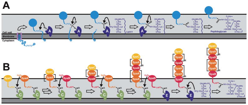
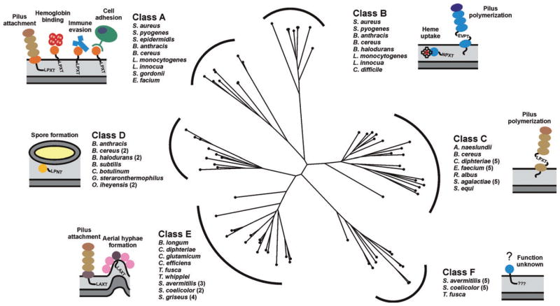
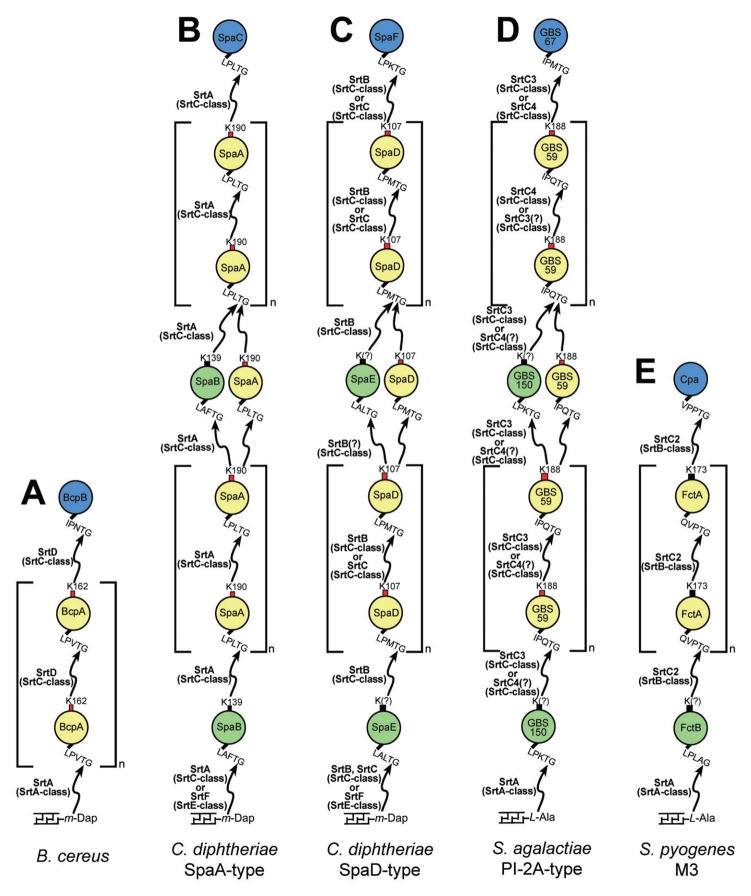
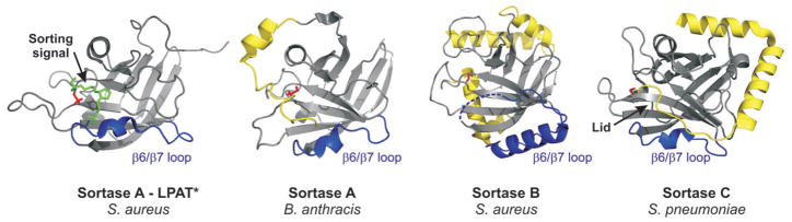
In Gram-positive bacteria proteins are displayed on the cell surface using sortase enzymes. These cysteine transpeptidases join proteins bearing an appropriate sorting signal to strategically positioned amino groups on the cell surface. Working alone, or in concert with other enzymes, sortases either attach proteins to the cross-bridge peptide of the cell wall or they link proteins together to form pili. Because surface proteins play a fundamental role in microbial physiology and are frequently virulence factors, sortase enzymes have been intensely studied since their discovery a little more than a decade ago. Based on their primary sequences and functions sortases can be partitioned into distinct families called class A to F enzymes. Most bacteria elaborate their surfaces using more than one type of sortase that function non-redundantly by recognizing unique sorting signals within their protein substrates. Here we review what is known about the functions of these enzymes and the molecular basis of catalysis. Particular emphasis is placed on 'pilin' specific class C sortases that construct structurally complex pili. Exciting new data have revealed that these enzymes are amazingly promiscuous in the substrates that they can employ and that there is a startling degree of diversity in their mechanism of action. We also review recent data that suggest that sortases are targeted to specific sites on the cell surface where they work with other sortases and accessory factors to properly function.


Figures

Fig. 1
Fig. 1
Mechanisms of sortase mediated attachment of surface proteins and pilus assembly at the bacterial cell wall. A. The S. aureus housekeeping sortase A anchors surface proteins to the peptidoglycan. The precursor protein containing an amino terminal leader peptide is secreted across the membrane through the Sec pathway. The exported protein (light blue) is processed by the sortase enzyme (dark blue, labelled ‘A’), which recognizes the LPXTG sequence and cleaves the surface proteins between the threonine and glycine residues of the motif. The enzyme then recognizes the pentaglycine cross-bridge peptide of lipid II as the second substrate. Subsequent formation of a peptide bond between the carbonyl of the threonine and the free amino group of the cross-bridge peptide results in covalent attachment of the protein to lipid II. The surface protein is then fully incorporated into the cross-linked peptidoglycan via the transglycosylation and transpeptidation reactions during the bacterial cell wall synthesis. The sphere coloured light blue represents the folded form of the cell surface displayed protein. B. Pilin-specific and housekeeping sortases assemble the SpaA pilus in C. diphtheria. The formation of complexes between the pilus-specific sortase C (light green) and the tip protein SpaC (light orange) initiates pilus assembly. The class C enzyme also recognizes the main pilin subunit SpaA (orange) forming SrtC–SpaA complexes. Nucleophilic attack by the free amino group originating from a lysine residue present in SpaA results in dissolution of the sortase–SpaC intermediate and the formation of a sortase–SpaA–SpaC complex. Repetition of this transpeptidation reaction results in pilus elongation. The class C sortase also incorporates the minor pilin SpaB (red) into the growing shaft by an analogous mechanism. Termination of pilus biogenesis is presumably initiated when the pilin polymer is transferred to the class E type housekeeping sortase (dark blue), which subsequently catalyses the nucleophilic attack by the amino group within lipid II. In the final assembly step the lipid II linked pilus is incorporated in the murein sacculus via normal cell wall biosynthesis.
Fig. 2
Fig. 2
Phylogenic tree showing the relationships among the six classes of sortases from Gram-positive bacteria. A multiple sequence alignment based on pairwise constraints of a selected set of 73 sortase proteins was generated using the program COBALT and a phylogenetic tree constructed using the neighbour joining method (Papadopoulos and Agarwala, 2007). The analysed sortases can be partitioned into six distinct subfamilies based on their primary sequences. It should be noted that the class D and E enzymes described here are collectively referred to as a class D enzymes by Bierne and colleagues (Dramsi et al., 2005). Class D and E enzymes have also previously been referred to as subfamily-4 and -5 enzymes (Comfort and Clubb, 2004). The bacterial species associated with the enzyme classes A–F are listed and schematic representations of the main biological function of their corresponding sortase substrates are illustrated. The bacterial species and accession numbers of the amino acid sequences used for the alignment are as followed: Actionomyces naeslundii (AAC13546), Bacillus anthracis (NP_847260; NP_843215; NP_846988), Bacillus cereus (NP_834511; NP_830752; NP_830495; NP_834250; NP_832268), Bacillus halodurans (NP_244463; NP_244878; NP_244160), Bacillus subtilis (NP_388801), Bifidobacterium longum (NP_695779), Clostridium botulinum (YP_001254630), Clostridium difficile (YP_001089230), Corynebacterium diphtheriae (NP_940343; NP_940532; NP_940531; NP_938627;NP_940575; NP_938624), Corynebacterium efficiens (NP_739396), Corynebacterium glutamicum (NP_602126), Enterococcus faecium (ZP_05713110; ZP_05714716; ZP_05713995; ZP_05713369; ZP_00603528; ZP_05713288), Geobacillus sp. (YP_003672707), Listeria innocua (NP_470268; NP_471617), Listeria monocytogenes (NP_464454; NP_465705), Oceanobacillus iheyensis (NP_691253; NP_694114), Ruminococcus albus (ZP_06717336), Streptomyces avermitilis (NP_826393; NP_827770; NP_826840; NP_828474; NP_821266; NP_825513; NP_825514; NP_826383; NP_825510), Streptomyces coelicolor (NP_628037; NP_628038; NP_627928; NP_631498; NP_626722; NP_625233; NP_627070), Streptomyces griseus (YP_001825232; YP_001825235; YP_001826193; YP_001825236), Staphylococcus aureus (NP_375640; NP_374252), Staphylococcus epidermis (NP_765631), Streptococcus agalactiae (NP_687667; NP_687668; NP_687670; NP_688403; NP_688404), Streptococcus equi (YP_002746265), Streptococcus gordonii (YP_001450517), Streptococcus pyogenes (NP_802272; NP_801365), Thermobifida fusca (YP_290439; YP_288977), Tropheryma whipplei (NP_787692).
Fig. 3
Fig. 3
The role of sortase enzymes in pilus assembly in various Gram-positive bacteria. Gram-positive bacteria are diverse in their assembly of pili with respect to the number of pilin subunits, number of sortases, class of sortases, as well as the type of sorting signals, lysine residues and cell wall acceptors recognized. Red boxes indicate lysine residues within a WXXXVXVYPK pilin motif, while black boxes indicate the absence of an obvious pilin motif. Lack of biochemical data demonstrating which lysine residue is involved in pilus polymerization is indicated with (?).
Fig. 4
Fig. 4
Sortase structure overview. Representative structures of sortase enzymes from different classes are shown. Class A: SrtA from S. aureus in complex with the sorting signal analogue LPAT* (green) (PDB code 2KID) and SrtA from B. anthracis (PDB code 2KW8). Class B: SrtB from S. aureus (PDB code 1NG5). Class C: SrtC1 from S. pneumoniae (PDB code 3G66). The β6/β7 loops are coloured in blue. Unique structural features are highlighted in yellow: the N-terminal extension in B. anthracis SrtA, the additional helices in SrtB from S. aureus and the flexible lid in S. pneumoniae SrtC1. The catalytic cysteine residues are highlighted in red and shown in a stick representation.

References

    1. Bagnoli F, Moschioni M, Donati C, Dimitrovska V, Ferlenghi I, Facciotti C, et al. A second pilus type in Streptococcus pneumoniae is prevalent in emerging serotypes and mediates adhesion to host cells. J Bacteriol. 2008;190:5480–5492. - PMC - PubMed
    1. Barnett TC, Patel AR, Scott JR. A novel sortase, SrtC2, from Streptococcus pyogenes anchors a surface protein containing a QVPTGV motif to the cell wall. J Bacteriol. 2004;186:5865–5875. - PMC - PubMed
    1. Bentley ML, Gaweska H, Kielec JM, McCafferty DG. Engineering the substrate specificity of Staphylococcus aureus Sortase A. The beta6/beta7 loop from SrtB confers NPQTN recognition to SrtA. J Biol Chem. 2007;282:6571–6581. - PubMed
    1. Boekhorst J, de Been MW, Kleerebezem M, Siezen RJ. Genome-wide detection and analysis of cell wall-bound proteins with LPxTG-like sorting motifs. J Bacteriol. 2005;187:4928–4934. - PMC - PubMed
    1. Budzik JM, Marraffini LA, Schneewind O. Assembly of pili on the surface of Bacillus cereus vegetative cells. Mol Microbiol. 2007;66:495–510. - PubMed

Publication types

MeSH terms

LinkOut - more resources