. 2011 Oct 25;2(6):e00168-11.
doi: 10.1128/mBio.00168-11. Print 2011.

Novel vaginal microflora colonization model providing new insight into microbicide mechanism of action

Affiliations

Novel vaginal microflora colonization model providing new insight into microbicide mechanism of action

Raina N Fichorova et al. mBio. .

Abstract

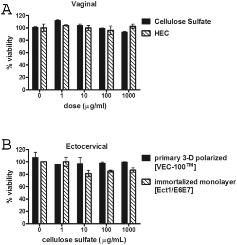
Several broad-spectrum microbicides, including cellulose sulfate (CS), have passed conventional preclinical and phase I clinical safety evaluation and yet have failed to protect women from acquiring HIV-1 in phase II/III trials. Concerns have been raised that current preclinical algorithms are deficient in addressing the complexity of the microflora-regulated vaginal mucosal barrier. We applied a novel microflora-colonized model to evaluate CS and hydroxyethylcellulose (HEC), which is used as a "universal placebo" in microbicide trials. Cervicovaginal epithelial cultures were colonized with normal vaginal microflora isolates representing common Lactobacillus species used as probiotics (L. acidophilus and L. crispatus) or Prevotella bivia and Atopobium vaginae, most prevalent in the disturbed microflora of bacterial vaginosis (BV). At baseline, all strains maintained constant epithelium-associated CFUs without inducing cytotoxicity and apoptosis. CS selectively reduced epithelium-associated CFUs and (to a lesser extent) planktonic CFUs, most significantly affecting L. crispatus. Inducing only minor changes in sterile epithelial cultures, CS induced expression of innate immunity mediators (RANTES, interleukin-8 [IL-8], and secretory leukocyte protease inhibitor [SLPI]) in microflora-colonized epithelia, most significantly potentiating effects of bacteria causing BV. In the absence of CS, all bacterial strains except L. acidophilus activated NF-κB, although IL-8 and RANTES levels were increased by the presence of BV-causing bacteria only. CS enhanced NF-κB activation in a dose-dependent manner under all conditions, including L. acidophilus colonization. HEC remained inert. These results offer insights into possible mechanisms of CS clinical failure. The bacterially colonized cervicovaginal model reveals unique aspects of microflora-epithelium-drug interactions and innate immunity in the female genital tract and should become an integral part of preclinical safety evaluation of anti-HIV microbicides and other vaginal formulations.

Importance: This report provides experimental evidence supporting the concept that the vaginal microflora regulates the epithelial innate immunity in a species- and strain-specific manner and that topically applied microbicides may alter both the bacterial and epithelial components of this homeostatic interaction. Our data also highlight the importance of differentiating the effects of biomedical interventions on epithelium-associated versus conventional planktonic bacterial growth when assessing vaginal mucosal health and immunity.


Figures

FIG 1
FIG 1
Colonization of human vaginal and cervical epithelial cells by vaginal bacteria showed consistent bacterial association with epithelial cells in the absence of apoptosis and cell toxicity. (A–D) Transmission electron microscopy image showing L. crispatus (A and B) and P. bivia (C and D) bacteria, visualized as electron-dense bodies, adherent to the surface of vaginal epithelial cells (Vk2/E6E7) with intact morphology after 24 h of colonization. The bars and images represent 500 nm and ×4,800 magnification (A), 500 nm and ×6,800 magnification (B), 2 µm and ×1,900 magnification (C), and 500 nm and ×13,000 magnification, respectively. (E) Caspase-3 cleavage is presented as percentages of cleaved total caspase-3 measured in vaginal epithelial cell lysates at 24 h after bacterial colonization or treatment with 1 µM staurosporine. Bars represent means and standard errors of the means (SEM) of the results determined with duplicate cultures used in two experiments. (F) Viability of vaginal epithelial cells assessed by trypan blue inclusion tests at 5 days postcolonization. Bars represent means and SEM of the results from triplicate culture experiments. (G) CFU counts per square centimeter of epithelial cell surface at 24 h and 48 h postcolonization of Vk2/E6E7 cells. Bars represent means and SEM of the results determined with triplicate cultures used in three experiments. (H) Parallel assessment of CFU counts associated with primary polarized (VEC-100) and immortalized monolayer (Ect1/E6E7) ectocervical epithelial cells at 48 h postcolonization.
FIG 2
FIG 2
Proinflammatory properties of vaginal microflora strains at 24 h postcolonization of epithelial monolayers in the absence of CS and HEC. (A) NF-κB activation assessed by luciferase activity. Bars represent means and SEM of the results determined with quadruplicate cultures in one of three independent experiments. (B) IL-8 levels measured in the vaginal epithelial cell culture supernatants. Bars represent means of the results determined with duplicate cultures in one of three experiments. For comparisons of the results determined with bacterially colonized cultures to those determined with control cultures without bacteria, ++ represents P < 0.01 and +++ represents P < 0.001 (ANOVA [Dunnett’s multiple-comparison test]).
FIG 3
FIG 3
Cell viability determined by the MTT assay after 24 h of compound exposure. (A) Immortalized vaginal epithelial cell monolayers (Vk2/E6E7) exposed to the same dose range of cellulose sulfate and hydroxyethylcellulose; (B) comparison of primary polarized (VEC-100) and immortalized monolayer (Ect1/E6E7) ectocervical epithelial cells exposed to cellulose sulfate. Values represent means and SEM of the results determined with duplicate cultures in one of three experiments.
FIG 4
FIG 4
Effects of cellulose sulfate and hydroxyethylcellulose (HEC) on bacterial colonization assessed after 24 h bacterial-epithelial coculture followed by 24h exposure to compound test doses. Bars represent means and SEM of CFUs associated with duplicate vaginal epithelial cell cultures in two experiments with two different batches of each test compound. For comparisons of the results determined with various compound doses to those determined with control medium, * represents P < 0.05, ** represents P < 0.01, and *** represents P < 0.001 (ANOVA [Dunnett’s multiple-comparison test]).
FIG 5
FIG 5
Direct effects of hydroxyethylcellulose (HEC) and cellulose sulfate on planktonic bacterial growth in the absence of epithelial cells. The bacterial suspensions that were used for epithelial colonization were simultaneously mixed with equal volumes of compound doses and incubated under anaerobic conditions for 24 h, followed by agar plating for enumeration of CFUs. Note that A. vaginae did not survive well in the absence of epithelial cells, whereas it maintained a stable colonization rate in the vaginal and cervical epithelial cells, as shown in Fig. 1G and H and 4D. Bars represent means and SEM of the results determined with triplicate cultures in two independent experiments performed with two different batches of test compounds. For comparisons of the results determined with various compound doses to those determined with control medium, *** represents P < 0.001 (ANOVA [Dunnett’s multiple-comparison test]).
FIG 6
FIG 6
Compound-induced innate immune responses after 24 h of colonization with vaginal bacterial strains followed by 24 h of exposure to cellulose sulfate or hydroxyethylcellulose. Bars represent means and SEM of RANTES (AB), IL-8 (CD), and SLPI (EF) levels measured in supernatants from duplicate ectocervical epithelial cell cultures in one of three experiments with three different batches of cellulose sulfate. The dashed line represents medium-control basal levels in the absence of bacterial colonization. Similar results were obtained with the vaginal epithelial cells (data not shown). For comparisons of the results determined with various compound doses to those determined with control medium within each treatment group (shown at the bottom), * represents P < 0.05 and ** represents P < 0.01; for comparisons of the results determined with bacterially colonized cultures to those determined with cultures without bacteria for each compound dose, + represents P < 0.05 and ++ represents P<0.01 (ANOVA [Dunnett’s multiple-comparison test]).
FIG 7
FIG 7
Compound-induced NF-κB activation after 24 h of bacterial colonization followed by 24 h of exposure to cellulose sulfate (A) or hydroxyethylcellulose (B). Bars represent means and SEM from luciferase activity determinations performed with duplicate cultures in one of three experiments with three different batches of each test compound. For comparisons of the results determined with various compound doses to those determined with control medium within each treatment group (shown at the right), ** represents P < 0.01; for comparisons of the results determined with bacterially colonized cultures to those determined with cultures without bacteria for each compound dose, ++ represents P < 0.01 and +++ represents P < 0.001 (ANOVA [Dunnett’s multiple-comparison test]).

References

    1. Fichorova RN, Anderson DJ. 1999. Differential expression of immunobiological mediators by immortalized human cervical and vaginal epithelial cells. Biol. Reprod. 60:508–514 - PubMed
    1. Fichorova RN, Cronin AO, Lien E, Anderson DJ, Ingalls RR. 2002. Response to Neisseria gonorrhoeae by cervicovaginal epithelial cells occurs in the absence of Toll-like receptor 4-mediated signaling. J. Immunol. 168:2424–2432 - PubMed
    1. Fichorova RN, Desai PJ, Gibson FC, III, Genco CA. 2001. Distinct proinflammatory host responses to Neisseria gonorrhoeae infection in immortalized human cervical and vaginal epithelial cells. Infect. Immun. 69:5840–5848 - PMC - PubMed
    1. Fichorova RN, et al. 2006. Trichomonas vaginalis lipophosphoglycan triggers a selective upregulation of cytokines by human female reproductive tract epithelial cells. Infect. Immun. 74:5773–5779 - PMC - PubMed
    1. Wira CR, Fahey JV, Sentman CL, Pioli PA, Shen L. 2005. Innate and adaptive immunity in female genital tract: cellular responses and interactions. Immunol. Rev. 206:306–335 - PubMed

Publication types