. 2011 Nov 8;108(45):18500-5.
doi: 10.1073/pnas.1103300108. Epub 2011 Oct 25.

A cardiac pathway of cyclic GMP-independent signaling of guanylyl cyclase A, the receptor for atrial natriuretic peptide

Affiliations

A cardiac pathway of cyclic GMP-independent signaling of guanylyl cyclase A, the receptor for atrial natriuretic peptide

Michael Klaiber et al. Proc Natl Acad Sci U S A. .

Abstract

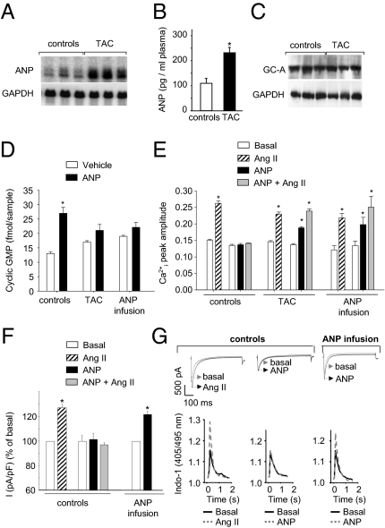
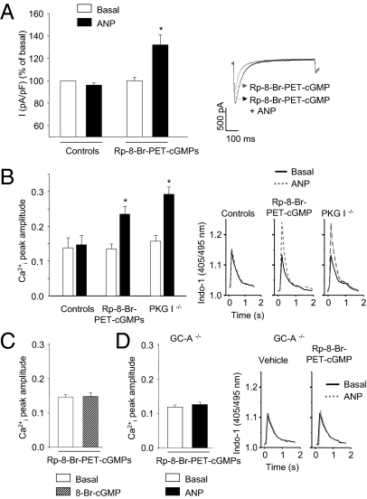
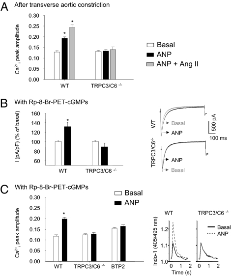
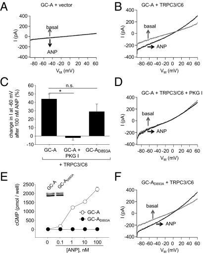
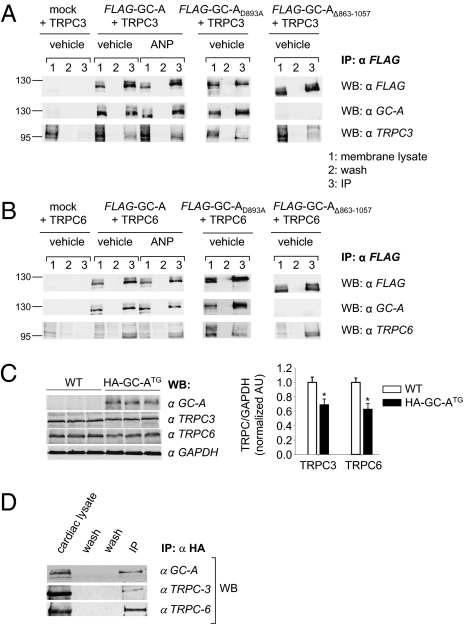
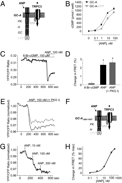
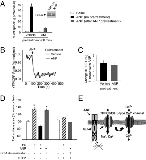
Cardiac atrial natriuretic peptide (ANP) regulates arterial blood pressure, moderates cardiomyocyte growth, and stimulates angiogenesis and metabolism. ANP binds to the transmembrane guanylyl cyclase (GC) receptor, GC-A, to exert its diverse functions. This process involves a cGMP-dependent signaling pathway preventing pathological [Ca(2+)](i) increases in myocytes. In chronic cardiac hypertrophy, however, ANP levels are markedly increased and GC-A/cGMP responses to ANP are blunted due to receptor desensitization. Here we show that, in this situation, ANP binding to GC-A stimulates a unique cGMP-independent signaling pathway in cardiac myocytes, resulting in pathologically elevated intracellular Ca(2+) levels. This pathway involves the activation of Ca(2+)-permeable transient receptor potential canonical 3/6 (TRPC3/C6) cation channels by GC-A, which forms a stable complex with TRPC3/C6 channels. Our results indicate that the resulting cation influx activates voltage-dependent L-type Ca(2+) channels and ultimately increases myocyte Ca(2)(+)(i) levels. These observations reveal a dual role of the ANP/GC-A-signaling pathway in the regulation of cardiac myocyte Ca(2+)(i) homeostasis. Under physiological conditions, activation of a cGMP-dependent pathway moderates the Ca(2+)(i)-enhancing action of hypertrophic factors such as angiotensin II. By contrast, a cGMP-independent pathway predominates under pathophysiological conditions when GC-A is desensitized by high ANP levels. The concomitant rise in [Ca(2+)](i) might increase the propensity to cardiac hypertrophy and arrhythmias.


Conflict of interest statement

The authors declare no conflict of interest.

Figures

Fig. 1.
Fig. 1.
Cardiac hypertrophy after transverse aortic constriction (TAC) is accompanied by altered myocyte cGMP and Ca2+ responses to ANP. (A) Northern blot showing cardiac expression of ANP and GAPDH. (B) Plasma levels of immunoreactive ANP (*P < 0.05 vs. controls). (C) Western blots showing levels of GC-A and GAPDH in lungs. (D) Effect of synthetic ANP (100 nM, 10 min) on cGMP contents of freshly isolated myocytes (*P < 0.05 vs. vehicle). (E and F) Peak amplitudes of Ca2+i transients [Indo-1 ratio405/495nm, systolic–diastolic (E)] and LTCC current densities [I (pA/pF), in percent (F)] at baseline and after application of Ang II, ANP, or ANP and Ang II (n = 6\x{2013}8; *P < 0.05 vs. basal). (G) LTCC current (Upper) and Ca2+i-transient traces (Lower) of three cells (from control vs. ANP-infused mice) at baseline and during superfusion with Ang II or ANP.
Fig. 2.
Fig. 2.
ANP, via GC-A signaling, increases LTCC currents and Ca2+i transients in myocytes with inhibited PKG I. Effect of ANP on LTCC current densities [I (pA/pF), in percent (A)] and Ca2+i transients (B) in untreated myocytes (controls), myocytes pretreated with the PKG I inhibitor Rp-8-Br-PET-cGMPs, and in PKG I-deficient (PKG I−/−) myocytes. The stimulatory effect of ANP on Ca2+i transients was not mimicked by 8-Br-cGMP, (C) and it was abolished in GC-A–deficient (GC-A−/−) myocytes (D). n = 6–8; *P < 0.05 vs. basal. Right panels in A, B, and D are examples of original tracings.
Fig. 3.
Fig. 3.
TRPC3/C6 channels mediate the Ca2+-stimulating effects of ANP in myocytes with inhibited cGMP/PKG I signaling. (A) Myocytes isolated from mice after TAC. Shown are peak amplitudes of Ca2+i transients at baseline and after application of ANP or ANP and Ang II in WT and TRPC3/C6−/− myocytes. (B and C) Myocytes pretreated with the PKG I inhibitor Rp-8-Br-PET-cGMPs. The effect of ANP on LTCC current densities [I (pA/pF), in percent (B)] and Ca2+i transients (C) of WT or TRPC3/C6−/− myocytes is shown. In addition, the effect of the TRPC channel inhibitor BTP2 on Ca2+i transients was tested (C). n = 6; *P < 0.05 vs. basal. Right panels in B and C are examples of original tracings.
Fig. 4.
Fig. 4.
ANP/GC-A stimulates TRPC3/C6 channel activity in transfected HEK293 cells. Shown are cation currents in HEK coexpressing GC-A and either vector [(A) n = 5] or TRPC3/C6 (BD) or TRPC3/C6 and PKG I (C and D) (all n = 10–13) and HEK coexpressing GC-AD893A and TRPC3/C6 (D and F) (n = 7). Cells additionally expressed EGFP and were identified by their marked fluorescence. Currents were recorded first under superfusion with bath solution (basal) and afterward during addition of 100 nM ANP. (A, B, D, and F) Current amplitudes in relation to membrane potential of representative cells and mean percentage of changes in response to ANP (C) (*P < 0.05). (E) Effects of ANP on cGMP levels in HEK expressing WT GC-A or GC-AD893A (n = 8). (Inset) Western blotting of GC-A and GC-AD893A membrane expression levels (all 10 μg protein per lane).
Fig. 5.
Fig. 5.
Stable association of GC-A and TRPC3 or TRPC6 in HEK293 cells and native myocytes. Coimmunoprecipitation of TRPC3 (A) or TRPC6 (B) with FLAG-GC-A, FLAG-GC-AD893A, or FLAG-GC-AΔ863–1057 from membranes of cotransfected HEK. Membrane fractions and immunoprecipitates (IP) with anti-FLAG antibody were separated on 10% SDS/PAGE and blotted with antibodies against FLAG, GC-A, TRPC3, or TRPC6. Incubation of HEK cells with ANP did not change the amount of coimmunoprecipitated FLAG-GC-A-TRPC proteins. The molecular weight in kilodaltons is shown at the left of the blot. (C) Western blot demonstrates cardiac overexpression of HA-tagged GC-A in transgenic compared with WT mice. Levels of TRPC3 and TRPC6 were slightly diminished in the transgenics (n = 3; *P < 0.05 vs. WT). (D) Heart lysates from HA-GC-ATG mice were immunoprecipitated with anti-HA antibody. The immunoblots with antibodies against GC-A, TRPC3, or TRPC6 showed coimmunoprecipitation of both TRPC3 and TRPC6 protein with HA-GC-A. All are representative Western blots of three independent experiments. Inputs were 1/10–1/20 of the protein used for IP. Control IPs using IgG do not give similar reactions.
Fig. 6.
Fig. 6.
FRET reveals the rapid ANP-modulated rearrangement of the GC-A–TRPC3 complex in cotransfected HEK293 cells. (A and F) Schemes of C-terminal CFP-labeled GC-A or GC-AΔ863–1057 and TRPC3-YFP. (B) Effects of ANP on cGMP levels in HEK expressing GC-A or GC-A-CFP. (C) Representative ratiometric recording of single-cell FRET signals in HEK cotransfected with GC-A-CFP and TRPC3-YFP at baseline and during superfusion of 8-Br-cGMP and ANP. (D) Relative FRET changes (n = 12; *P < 0.05 vs. baseline). (E) Effects of ANP on FRET between GC-A-CFP and TRPC3-YFP in HEK additionally cotransfected with PKG I (n = 6). (G) Representative ratiometric recordings of single-cell FRET signals in HEK cotransfected with GC-AΔ863–1057-CFP and TRPC3-YFP at baseline and during superfusion with ANP. (H) Concentration-dependent effects of ANP on FRET between GC-AΔ863–1057-CFP and TRPC3-YFP (EC50 ∼30 nM; n = 11).
Fig. 7.
Fig. 7.
Homologous desensitization of GC-A impairs guanylyl cyclase activity but preserves the effect of ANP on rearrangement of the GC-A–TRPC3 complex, revealing prohypertrophic actions of the hormone. (A) GC-A expressing HEK293 cells were pretreated with vehicle or ANP (100 nM, 60 min) before preparation of crude cell membranes for determination of basal (vehicle) vs. acute ANP-stimulated cGMP formation (n = 5, *P < 0.05 vs. basal). (Inset) Western blotting of GC-A expression levels in vehicle vs. ANP-pretreated cells (all 10 μg protein per lane). (B) Recordings of single-cell FRET signals in HEK cotransfected with GC-A-CFP and TRPC3-YFP at baseline and in response to acute addition of ANP. (C) Relative FRET changes evoked by ANP in vehicle- or ANP-pretreated cells (n = 11). (D) Cell surface area of neonatal rat ventricular myocytes treated with phenylephrine (PE, 100 μM, 48 h) in the absence (−) or presence (+) of ANP (100 nM, 48 h) or with ANP (100 nM, 48 h) after GC-A desensitization (100 nM ANP, 60 min of pretreatment). Effect of BTP2 (2 μM, 15 min pretreatment) on growth responses to PE and ANP (n = 3; 100 cells per experiment). (E) Schematic model showing how GC-A associates with TRPC3/C6 channels to form a stable complex in myocytes. When cGMP formation by GC-A is blunted, ANP via GC-A directly activates TRPC3 and TRPC6 (TRPC3/C6) channels. Subsequent cation influx ultimately activates LTCC currents.

References

    1. Kuhn M. Structure, regulation, and function of mammalian membrane guanylyl cyclase receptors, with a focus on guanylyl cyclase-A. Circ Res. 2003;93:700–709. - PubMed
    1. Holtwick R, et al. Pressure-independent cardiac hypertrophy in mice with cardiomyocyte-restricted inactivation of the atrial natriuretic peptide receptor guanylyl cyclase-A. J Clin Invest. 2003;111:1399–1407. - PMC - PubMed
    1. Klaiber M, et al. Novel insights into the mechanisms mediating the local antihypertrophic effects of cardiac atrial natriuretic peptide: Role of cGMP-dependent protein kinase and RGS2. Basic Res Cardiol. 2010;105:583–595. - PMC - PubMed
    1. Kinoshita H, et al. Inhibition of TRPC6 channel activity contributes to the antihypertrophic effects of natriuretic peptides-guanylyl cyclase-A signaling in the heart. Circ Res. 2010;106:1849–1860. - PubMed
    1. Koitabashi N, et al. Cyclic GMP/PKG-dependent inhibition of TRPC6 channel activity and expression negatively regulates cardiomyocyte NFAT activation novel mechanism of cardiac stress modulation by PDE5 inhibition. J Mol Cell Cardiol. 2010;48:713–724. - PMC - PubMed

Publication types